5OAK image
Deposition Date 2017-06-22
Release Date 2018-02-21
Last Version Date 2024-01-17
Entry Detail
PDB ID:
5OAK
Keywords:
Title:
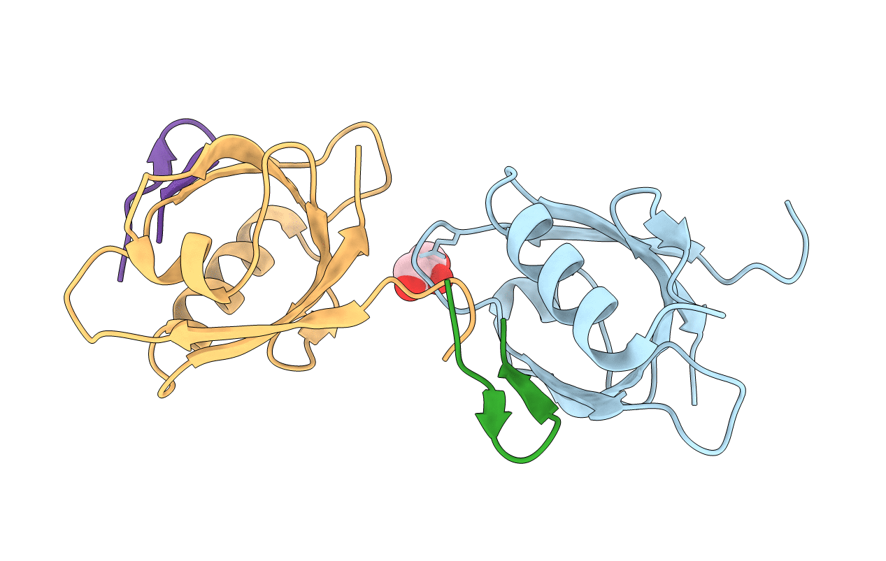
Structure of the dmPar3 PDZ1 domain in complex with the dmPar6 PBM
Biological Source:
Source Organism:
Host Organism:
Method Details:
Experimental Method:
Resolution:
1.50 Å
R-Value Free:
0.16
R-Value Work:
0.14
R-Value Observed:
0.14
Space Group:
P 31
Macromolecular Entities
Polymer Type:polypeptide(L)
Molecule:Bazooka, isoform C,LD29223p
Chain IDs:A, B, C, D
Chain Length:117
Number of Molecules:4
Biological Source:Drosophila melanogaster
Ligand Molecules
Primary Citation
Structural basis for the interaction between the cell polarity proteins Par3 and Par6.
Sci Signal 11 ? ? (2018)
PMID: 29440511 DOI: 10.1126/scisignal.aam9899

Abstact

Polarity is a fundamental property of most cell types. The Par protein complex is a major driving force in generating asymmetrically localized protein networks and consists of atypical protein kinase C (aPKC), Par3, and Par6. Dysfunction of this complex causes developmental abnormalities and diseases such as cancer. We identified a PDZ domain-binding motif in Par6 that was essential for its interaction with Par3 in vitro and for Par3-mediated membrane localization of Par6 in cultured cells. In fly embryos, we observed that the PDZ domain-binding motif was functionally redundant with the PDZ domain in targeting Par6 to the cortex of epithelial cells. Our structural analyses by x-ray crystallography and NMR spectroscopy showed that both the PDZ1 and PDZ3 domains but not the PDZ2 domain in Par3 engaged in a canonical interaction with the PDZ domain-binding motif in Par6. Par3 thus has the potential to recruit two Par6 proteins simultaneously, which may facilitate the assembly of polarity protein networks through multivalent PDZ domain interactions.

Legend

Protein

Chemical

Disease

Primary Citation of related structures