5O8X image
Deposition Date 2017-06-14
Release Date 2017-11-29
Last Version Date 2024-05-08
Entry Detail
PDB ID:
5O8X
Keywords:
Title:
The X-ray Structure of Catenated Lytic Transglycosylase SltB1 from Pseudomonas aeruginosa
Biological Source:
Source Organism:
Host Organism:
Method Details:
Experimental Method:
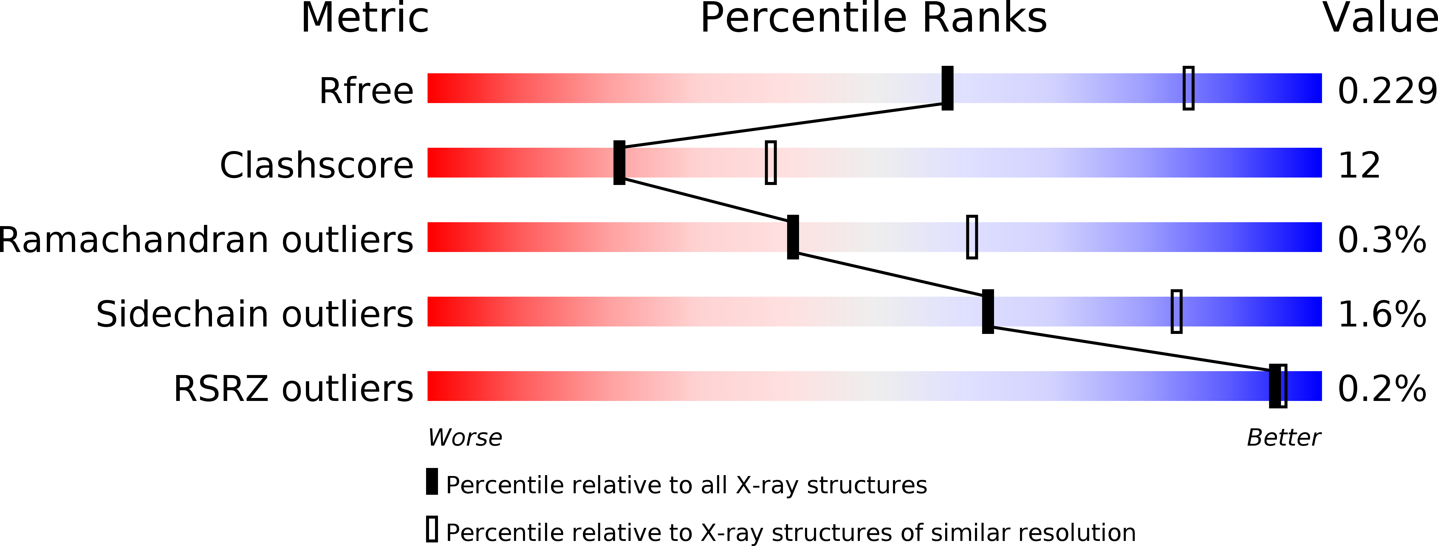
Resolution:
2.50 Å
R-Value Free:
0.22
R-Value Work:
0.17
R-Value Observed:
0.17
Space Group:
C 1 2 1
Macromolecular Entities
Structures with similar UniProt ID
Protein Blast
Polymer Type:polypeptide(L)
Molecule:Soluble lytic transglycosylase B
Chain IDs:A, B
Chain Length:306
Number of Molecules:2
Biological Source:Pseudomonas aeruginosa
Primary Citation
X-ray Structure of Catenated Lytic Transglycosylase SltB1.
Biochemistry 56 6317 6320 (2017)
PMID: 29131935 DOI: 10.1021/acs.biochem.7b00932

Abstact

Formation of catenanes by proteins is rare, with few known examples. We report herein the X-ray structure of a catenane dimer of lytic transglycosylase SltB1 of Pseudomonas aeruginosa. The enzyme is soluble and exists in the periplasmic space, where it modifies the bacterial cell wall. The catenane dimer exhibits the protein monomers in a noncovalent chain-link arrangement, whereby a stretch of 51 amino acids (to become a loop and three helices) from one monomer threads through the central opening of the structure of the partner monomer. The protein folds after threading in a manner that leaves two helices (α1 and α2) as stoppers to impart stability to the dimer structure. The symmetric embrace by the two SltB1 molecules occludes both active sites entirely, an arrangement that is sustained by six electrostatic interactions between the two monomers. In light of the observation of these structural motifs in all members of Family 3 lytic transglycosylases, catenanes might be present for those enzymes, as well. The dimeric catenane might represent a regulated form of SltB1.

Legend

Protein

Chemical

Disease

Primary Citation of related structures
Feedback Form
Name
Email
Institute
Feedback