
Deposition Date
2017-06-02
Release Date
2018-10-24
Last Version Date
2024-01-17
Entry Detail
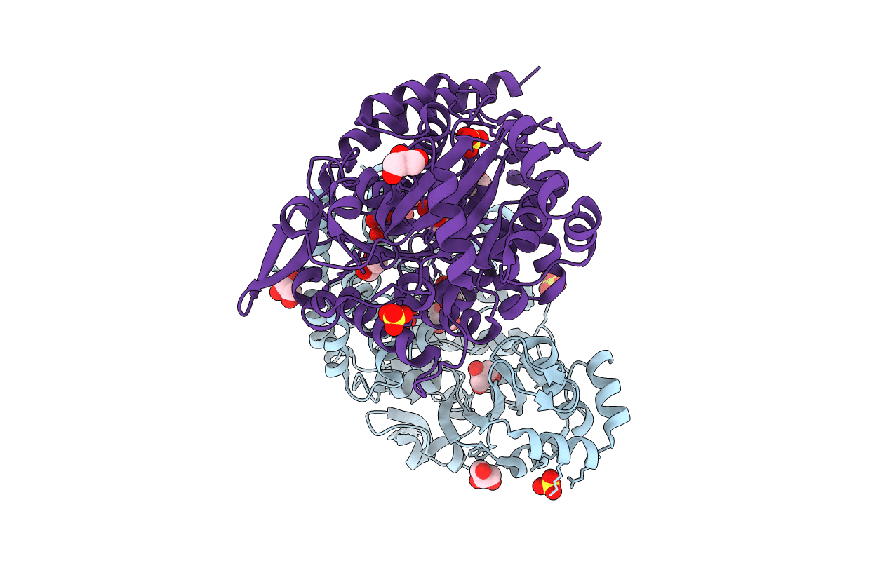
PDB ID:
5O5X
Keywords:
Title:
Crystal structure of Thermococcus litoralis ADP-dependent glucokinase (GK)
Biological Source:
Source Organism(s):
Expression System(s):
Method Details:
Experimental Method:
Resolution:
2.15 Å
R-Value Free:
0.22
R-Value Work:
0.19
R-Value Observed:
0.19
Space Group:
P 21 21 21
