5O07 image
Deposition Date 2017-05-16
Release Date 2017-08-02
Last Version Date 2024-01-17
Entry Detail
PDB ID:
5O07
Keywords:
Title:
The crystal structure of the human carbonic anhydrase II in complex with a nitroimidazole sulfamate inhibitor
Biological Source:
Source Organism:
Homo sapiens (Taxon ID: 9606)
Method Details:
Experimental Method:
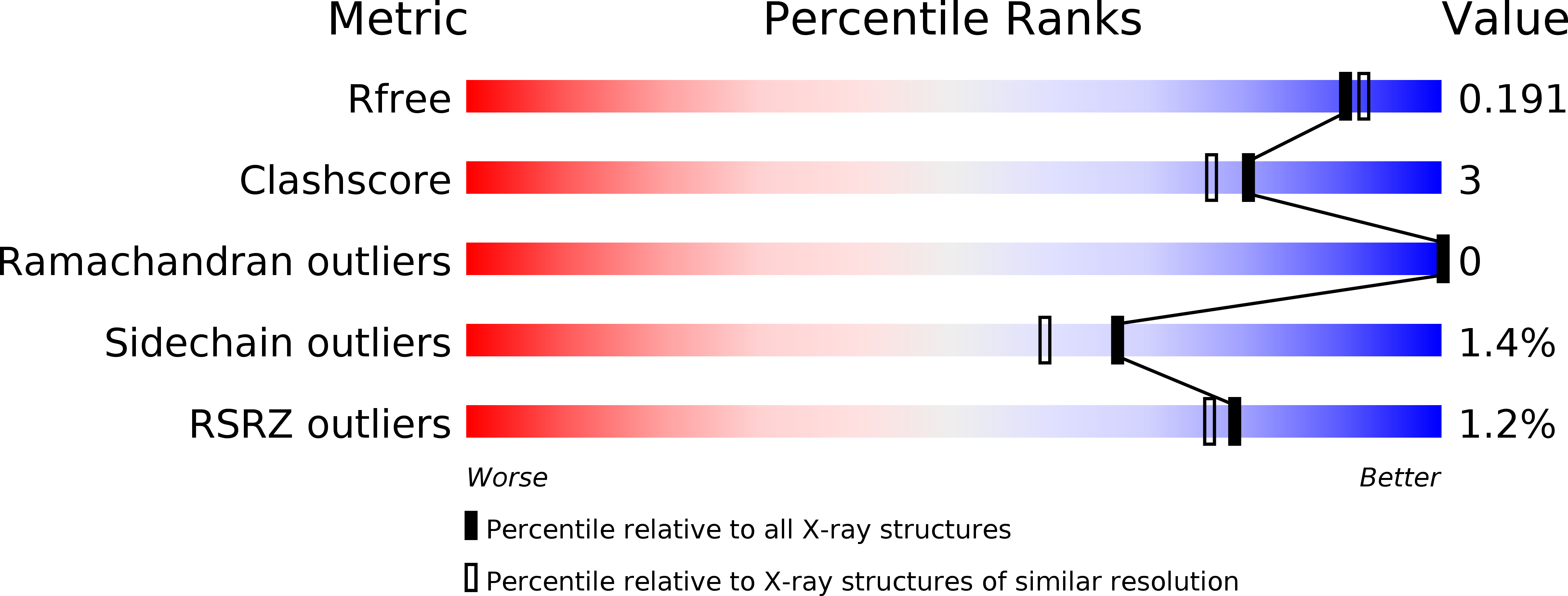
Resolution:
1.80 Å
R-Value Free:
0.19
R-Value Work:
0.15
Space Group:
P 1 21 1
Macromolecular Entities
Polymer Type:polypeptide(L)
Molecule:Carbonic anhydrase 2
Gene (Uniprot):CA2
Chain IDs:A
Chain Length:260
Number of Molecules:1
Biological Source:Homo sapiens
Primary Citation
Insights into the binding mode of sulphamates and sulphamides to hCA II: crystallographic studies and binding free energy calculations.
J Enzyme Inhib Med Chem 32 1002 1011 (2017)
PMID: 28738704 DOI: 10.1080/14756366.2017.1349764

Abstact

Sulphamate and sulphamide derivatives have been largely investigated as carbonic anhydrase inhibitors (CAIs) by means of different experimental techniques. However, the structural determinants responsible for their different binding mode to the enzyme active site were not clearly defined so far. In this paper, we report the X-ray crystal structure of hCA II in complex with a sulphamate inhibitor incorporating a nitroimidazole moiety. The comparison with the structure of hCA II in complex with its sulphamide analogue revealed that the two inhibitors adopt a completely different binding mode within the hCA II active site. Starting from these results, we performed a theoretical study on sulphamate and sulphamide derivatives, demonstrating that electrostatic interactions with residues within the enzyme active site play a key role in determining their binding conformation. These findings open new perspectives in the design of effective CAIs using the sulphamate and sulphamide zinc binding groups as lead compounds.

Legend

Protein

Chemical

Disease

Primary Citation of related structures