5NWX image
Deposition Date 2017-05-08
Release Date 2017-12-13
Last Version Date 2024-01-17
Entry Detail
PDB ID:
5NWX
Keywords:
Title:
Insight into the molecular recognition mechanism of the coactivator NCoA1 by STAT6
Biological Source:
Source Organism:
Mus musculus (Taxon ID: 10090)
Homo sapiens (Taxon ID: 9606)
Method Details:
Experimental Method:
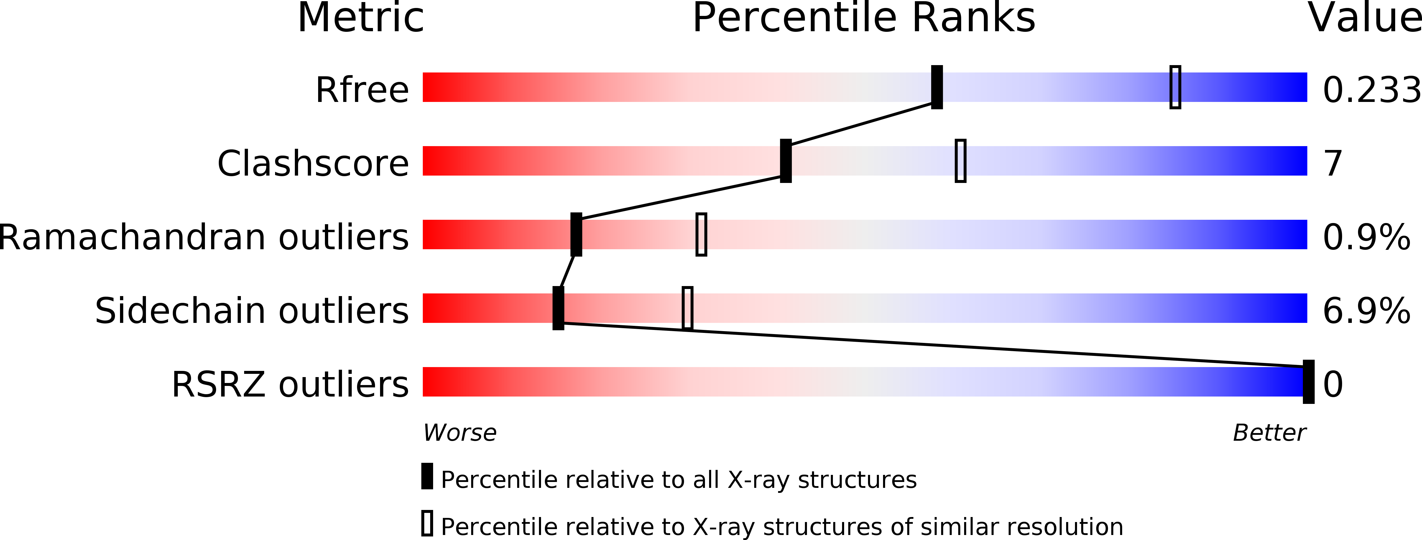
Resolution:
2.51 Å
R-Value Free:
0.23
R-Value Work:
0.18
R-Value Observed:
0.18
Space Group:
P 62
Macromolecular Entities
Structures with similar UniProt ID
Protein Blast
Polymer Type:polypeptide(L)
Molecule:Nuclear receptor coactivator 1
Gene (Uniprot):Ncoa1
Mutagens:K343R
Chain IDs:A
Chain Length:132
Number of Molecules:1
Biological Source:Mus musculus
Structures with similar UniProt ID
Protein Blast
Polymer Type:polypeptide(L)
Molecule:Signal transducer and activator of transcription 6
Gene (Uniprot):STAT6
Chain IDs:B
Chain Length:32
Number of Molecules:1
Biological Source:Homo sapiens
Primary Citation
Insight into the molecular recognition mechanism of the coactivator NCoA1 by STAT6.
Sci Rep 7 16845 16845 (2017)
PMID: 29203888 DOI: 10.1038/s41598-017-17088-5

Abstact

Crucial for immune and anti-inflammatory cellular responses, signal transducer and activator of transcription 6 (STAT6) regulates transcriptional activation in response to interleukin-4 and -13 -induced tyrosine phosphorylation by direct interaction with coactivators. The interaction of STAT6 with nuclear coactivator 1 (NCoA1) is mediated by a short region of the STAT6 transactivation domain that includes the motif LXXLL and interacts with the PAS-B domain of NCoA1. Despite the availability of an X-ray structure of the PAS-B domain/ Leu794-Gly814-STAT6 complex, the mechanistic details of this interaction are still poorly understood. Here, we determine the structure of the NCoA1257-385/STAT6783-814 complex using Nuclear Magnetic Resonance (NMR) and X-ray crystallography. The STAT6783-814 peptide binds with additional N-terminal amino acids to NCoA1257-385, compared to the STAT6794-814 peptide, explaining its higher affinity. Secondary and tertiary structures existing in the free peptide are more highly populated in the complex, suggesting binding by conformational selection.

Legend

Protein

Chemical

Disease

Primary Citation of related structures