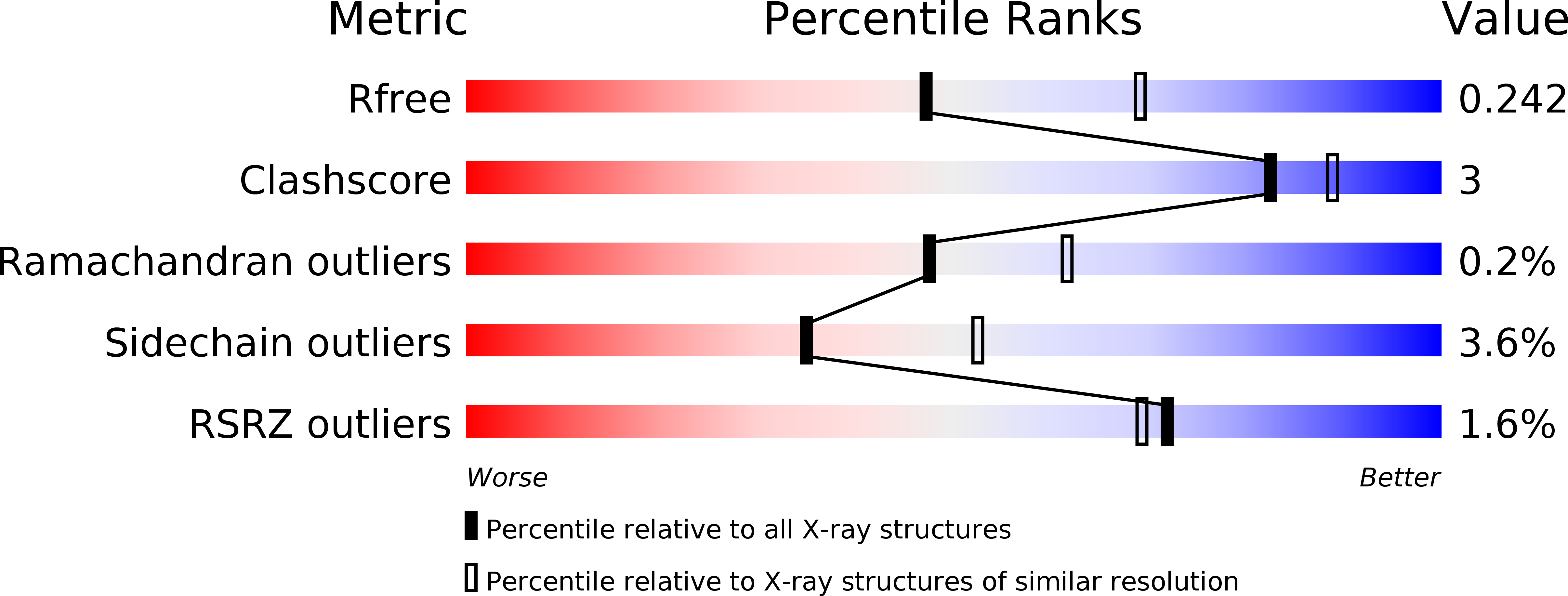
5NSF image
Deposition Date 2017-04-26
Release Date 2019-01-16
Last Version Date 2024-01-17
Entry Detail
PDB ID:
5NSF
Keywords:
Title:
Structure of AzuAla
Biological Source:
Method Details:
Experimental Method:
Resolution:
2.43 Å
R-Value Free:
0.23
R-Value Work:
0.18
R-Value Observed:
0.18
Space Group:
P 1
Macromolecular Entities
Structures with similar UniProt ID
Protein Blast
Polymer Type:polypeptide(L)
Molecule:Tyrosine--tRNA ligase
Gene (Uniprot):tyrS
Chain IDs:A (auth: B), B (auth: A), C, D
Chain Length:318
Number of Molecules:4
Biological Source:Methanocaldococcus jannaschii (strain ATCC 43067 / DSM 2661 / JAL-1 / JCM 10045 / NBRC 100440)
Primary Citation
Site-Resolved Observation of Vibrational Energy Transfer Using a Genetically Encoded Ultrafast Heater.
Angew. Chem. Int. Ed. Engl. 58 2899 2903 (2019)
PMID: 30589180 DOI: 10.1002/anie.201812995

Abstact

Allosteric information transfer in proteins has been linked to distinct vibrational energy transfer (VET) pathways in a number of theoretical studies. Experimental evidence for such pathways, however, is sparse because site-selective injection of vibrational energy into a protein, that is, localized heating, is required for their investigation. Here, we solved this problem by the site-specific incorporation of the non-canonical amino acid β-(1-azulenyl)-l-alanine (AzAla) through genetic code expansion. As an exception to Kasha's rule, AzAla undergoes ultrafast internal conversion and heating after S1 excitation while upon S2 excitation, it serves as a fluorescent label. We equipped PDZ3, a protein interaction domain of postsynaptic density protein 95, with this ultrafast heater at two distinct positions. We indeed observed VET from the incorporation sites in the protein to a bound peptide ligand on the picosecond timescale by ultrafast IR spectroscopy. This approach based on genetically encoded AzAla paves the way for detailed studies of VET and its role in a wide range of proteins.

Legend

Protein

Chemical

Disease

Primary Citation of related structures