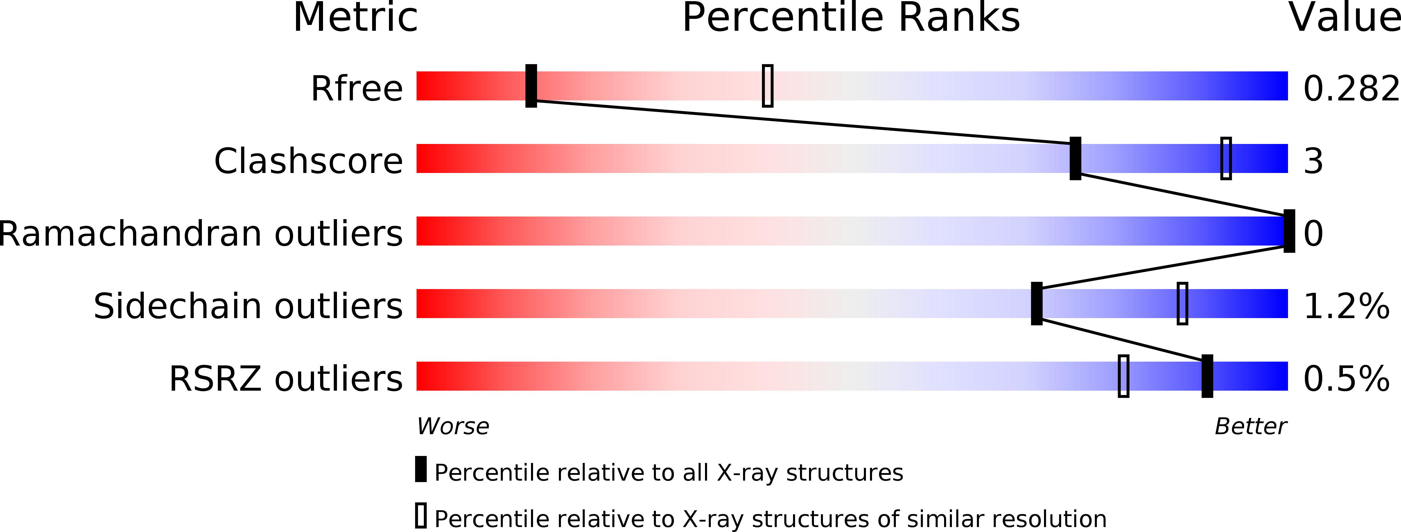
5NPV image
Deposition Date 2017-04-19
Release Date 2017-10-11
Last Version Date 2024-01-17
Entry Detail
PDB ID:
5NPV
Keywords:
Title:
Structure of human ATG5-ATG16L1(ATG5BD) complex (I4)
Biological Source:
Source Organism:
Homo sapiens (Taxon ID: 9606)
Host Organism:
Method Details:
Experimental Method:
Resolution:
3.10 Å
R-Value Free:
0.27
R-Value Work:
0.25
R-Value Observed:
0.25
Space Group:
I 4
Macromolecular Entities
Polymer Type:polypeptide(L)
Molecule:Autophagy protein 5
Gene (Uniprot):ATG5
Chain IDs:A, C
Chain Length:282
Number of Molecules:2
Biological Source:Homo sapiens
Polymer Type:polypeptide(L)
Molecule:Autophagy-related protein 16-1
Gene (Uniprot):ATG16L1
Chain IDs:B, D
Chain Length:301
Number of Molecules:2
Biological Source:Homo sapiens
Primary Citation
Identification, biochemical characterization and crystallization of the central region of human ATG16L1.
Acta Crystallogr F Struct Biol Commun 73 560 567 (2017)
PMID: 28994404 DOI: 10.1107/S2053230X17013280

Abstact

ATG16L1 plays a major role in autophagy. It acts as a molecular scaffold which mediates protein-protein interactions essential for autophagosome formation. The ATG12~ATG5-ATG16L1 complex is one of the key complexes involved in autophagosome formation. Human ATG16L1 comprises 607 amino acids with three functional domains named ATG5BD, CCD and WD40, where the C-terminal WD40 domain represents approximately 50% of the full-length protein. Previously, structures of the C-terminal WD40 domain of human ATG16L1 as well as of human ATG12~ATG5 in complex with the ATG5BD of ATG16L1 have been reported. However, apart from the ATG5BD, no structural information for the N-terminal half, including the CCD, of human ATG16L1 is available. In this study, the authors aimed to structurally characterize the N-terminal half of ATG16L1. ATG16L111-307 in complex with ATG5 has been purified and crystallized in two crystal forms. However, both crystal structures revealed degradation of ATG16L1, resulting in crystals comprising only full-length ATG5 and the ATG5BD of ATG16L1. The structures of ATG5-ATG5BD in two novel crystal forms are presented, further supporting the previously observed dimerization of ATG5-ATG16L1. The reported degradation points towards a high instability at the linker region between the ATG5BD and the CCD in ATG16L1. Based on this observation and further biochemical analysis of ATG16L1, a stable 236-amino-acid subfragment comprising residues 72-307 of the N-terminal half of ATG16L1, covering the residual, so far structurally uncharacterized region of human ATG16L1, was identified. Here, the identification, purification, biochemical characterization and crystallization of the proteolytically stable ATG16L172-307 subfragment are reported.

Legend

Protein

Chemical

Disease

Primary Citation of related structures