Deposition Date
2017-04-07
Release Date
2017-11-29
Last Version Date
2024-11-20
Entry Detail
PDB ID:
5NMT
Keywords:
Title:
Dimer structure of Sortilin ectodomain crystal form 1, 2.3A
Biological Source:
Source Organism(s):
Mus musculus (Taxon ID: 10090)
Expression System(s):
Method Details:
Experimental Method:
Resolution:
2.30 Å
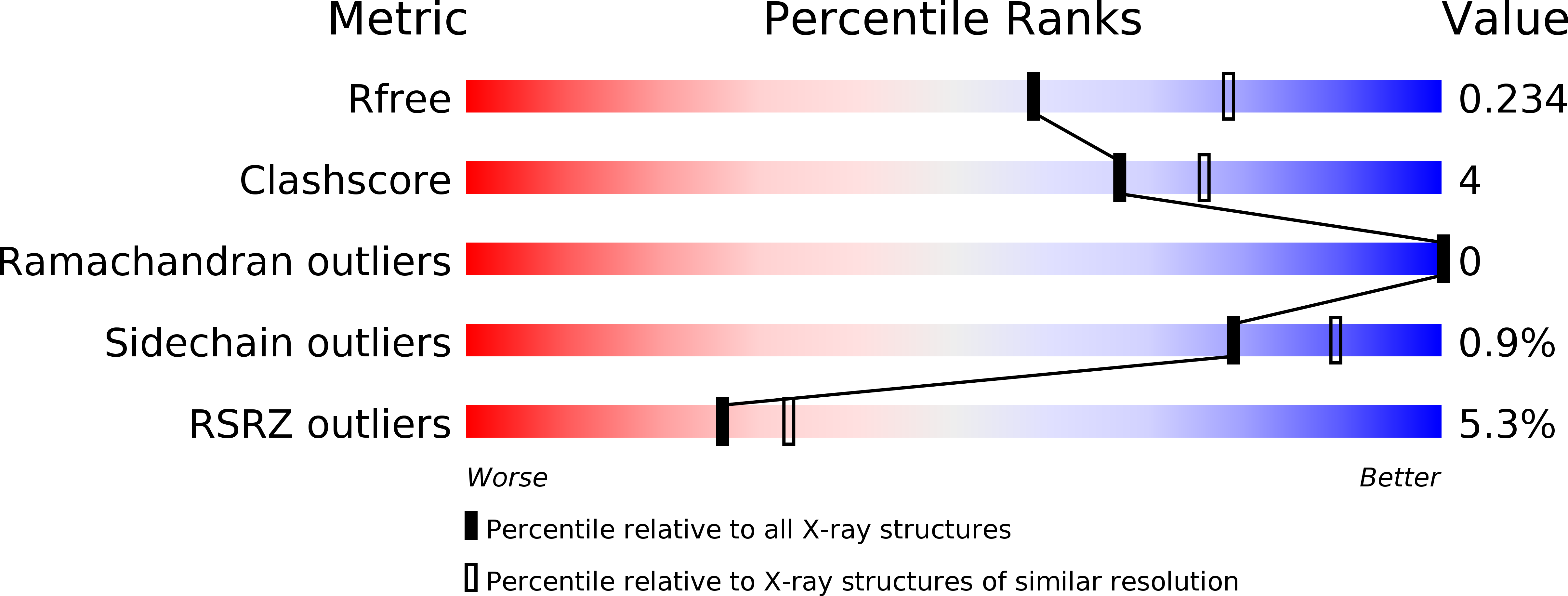
R-Value Free:
0.23
R-Value Work:
0.20
R-Value Observed:
0.20
Space Group:
P 21 21 21