Deposition Date
2017-03-24
Release Date
2017-12-06
Last Version Date
2024-01-17
Entry Detail
PDB ID:
5NIN
Keywords:
Title:
Crystal Structure of AKAP79 calmodulin binding domain peptide in complex with Ca2+/Calmodulin
Biological Source:
Source Organism(s):
Homo sapiens (Taxon ID: 9606)
Expression System(s):
Method Details:
Experimental Method:
Resolution:
1.70 Å
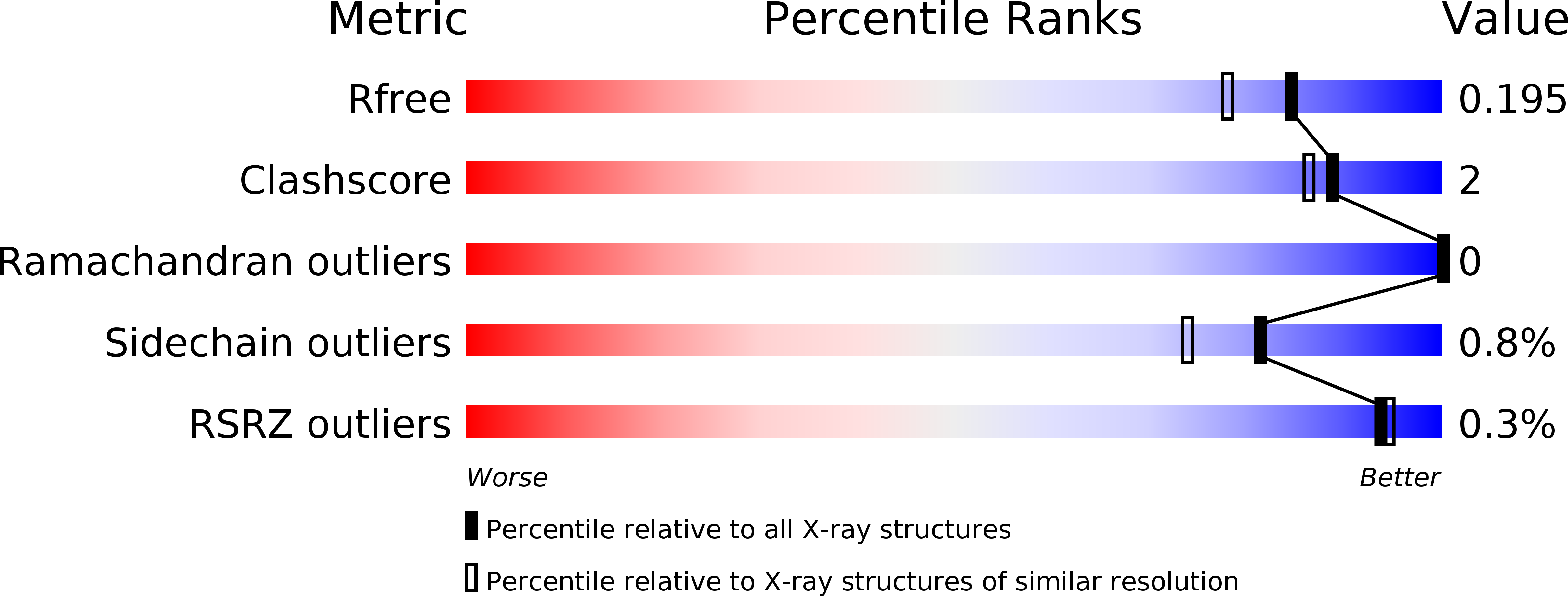
R-Value Free:
0.19
R-Value Work:
0.16
R-Value Observed:
0.16
Space Group:
P 42 21 2