
Deposition Date
2017-03-22
Release Date
2017-04-12
Last Version Date
2024-05-08
Entry Detail
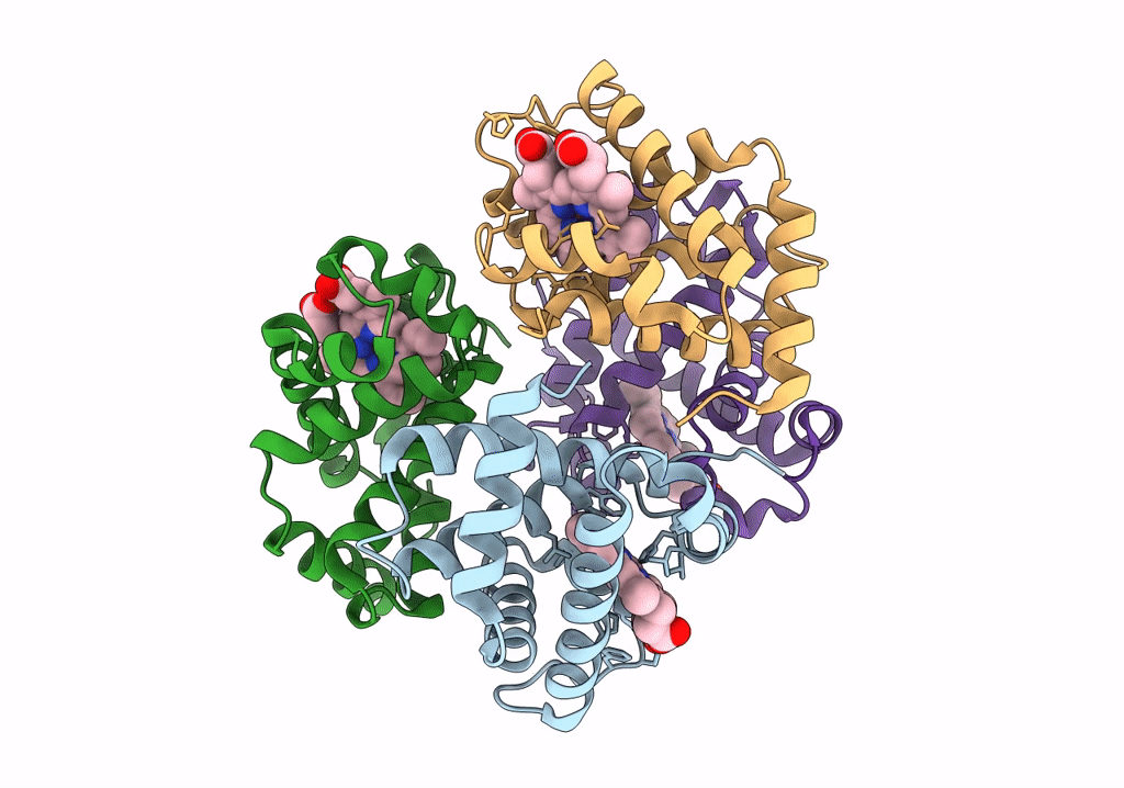
PDB ID:
5NI1
Keywords:
Title:
CryoEM structure of haemoglobin at 3.2 A determined with the Volta phase plate
Biological Source:
Source Organism(s):
Homo sapiens (Taxon ID: 9606)
Expression System(s):
Method Details:
Experimental Method:
Resolution:
3.20 Å
Aggregation State:
PARTICLE
Reconstruction Method:
SINGLE PARTICLE
