
Deposition Date
2017-03-11
Release Date
2017-06-21
Last Version Date
2024-10-16
Entry Detail
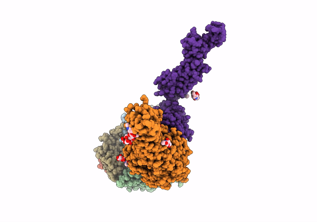
PDB ID:
5NET
Keywords:
Title:
Localised Reconstruction of Integrin alpha V beta 6 bound to Foot and Mouth Disease Virus O1 Manisa - Pose A.
Biological Source:
Source Organism(s):
Foot-and-mouth disease virus (Taxon ID: 12110)
Homo sapiens (Taxon ID: 9606)
Homo sapiens (Taxon ID: 9606)
Expression System(s):
Method Details:
Experimental Method:
Resolution:
8.60 Å
Aggregation State:
PARTICLE
Reconstruction Method:
SINGLE PARTICLE
