5NEP image
Deposition Date 2017-03-11
Release Date 2017-05-31
Last Version Date 2024-05-08
Entry Detail
PDB ID:
5NEP
Keywords:
Title:
The structure of the G. violaceus guanidine II riboswitch P1 stem-loop with methylguanidine
Biological Source:
Source Organism:
Method Details:
Experimental Method:
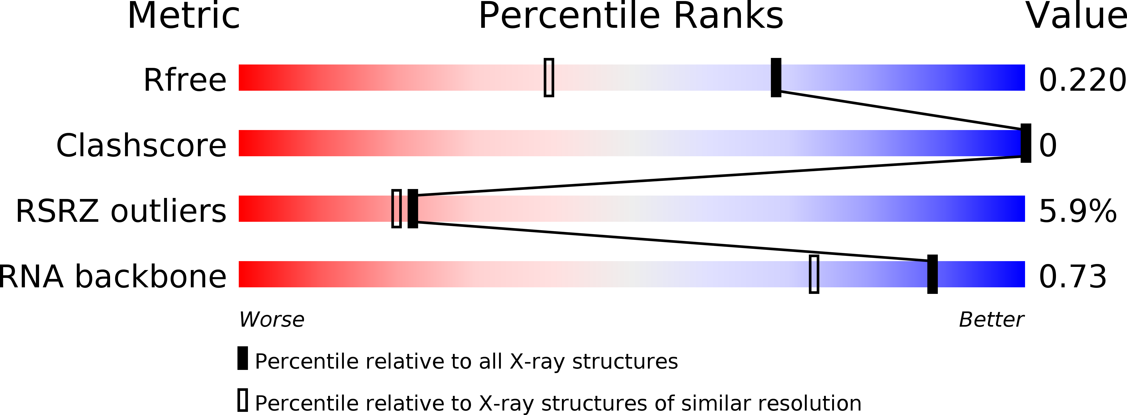
Resolution:
1.60 Å
R-Value Free:
0.21
R-Value Work:
0.20
R-Value Observed:
0.20
Space Group:
H 3 2
Macromolecular Entities
Polymer Type:polyribonucleotide
Molecule:RNA (5'-R(*GP*GP*UP*GP*GP*GP*GP*AP*CP*GP*AP*CP*CP*CP*CP*AP*(CBV)P*C)-3')
Chain IDs:A
Chain Length:18
Number of Molecules:1
Biological Source:Gloeobacter violaceus
Primary Citation
The Structure of the Guanidine-II Riboswitch.
Cell Chem Biol 24 695 702.e2 (2017)
PMID: 28529131 DOI: 10.1016/j.chembiol.2017.05.014

Abstact

The guanidine-II (mini-ykkC) riboswitch is the smallest of the guanidine-responsive riboswitches, comprising two stem loops of similar sequence. We have solved high-resolution crystal structures of both stem loops for the riboswitch from Gloeobacter violaceus. The stem loops have a strong propensity to dimerize by intimate loop-loop interaction. The dimerization creates specific binding pockets for two guanidine molecules, explaining their cooperative binding. Within the binding pockets the ligands are hydrogen bonded to a guanine at O6 and N7, and to successive backbone phosphates. Additionally they are each stacked upon a guanine nucleobase. One side of the pocket has an opening to the solvent, slightly lowering the specificity of ligand binding, and structures with bound methylguanidine, aminoguanidine, and agmatine show how this is possible.

Legend

Protein

Chemical

Disease

Primary Citation of related structures