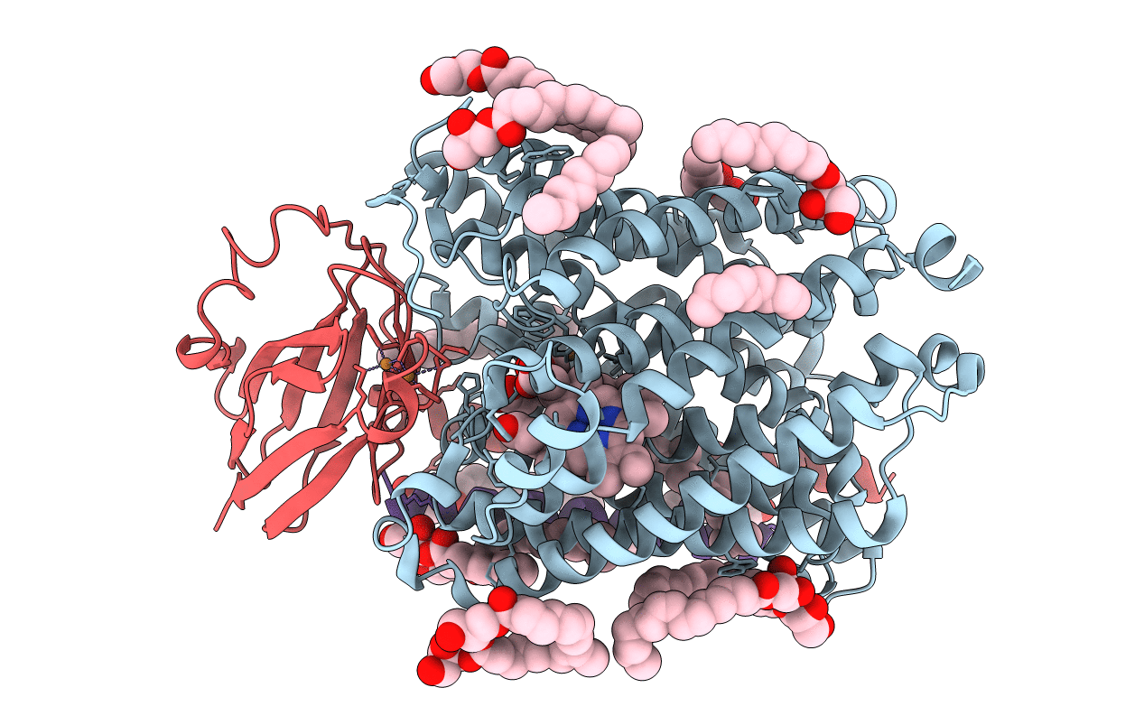
5NDC image
Deposition Date 2017-03-08
Release Date 2017-08-23
Last Version Date 2024-01-17
Entry Detail
PDB ID:
5NDC
Keywords:
Title:
Structure of ba3-type cytochrome c oxidase from Thermus thermophilus by serial femtosecond crystallography
Biological Source:
Source Organism:
Method Details:
Experimental Method:
Resolution:
2.30 Å
R-Value Free:
0.19
R-Value Work:
0.16
R-Value Observed:
0.16
Space Group:
C 1 2 1
Macromolecular Entities
Polymer Type:polypeptide(L)
Molecule:Cytochrome c oxidase subunit 1
Gene (Uniprot):cbaA
Chain IDs:A
Chain Length:569
Number of Molecules:1
Biological Source:Thermus thermophilus
Polymer Type:polypeptide(L)
Molecule:Cytochrome c oxidase subunit 2
Gene (Uniprot):cbaB
Chain IDs:B
Chain Length:168
Number of Molecules:1
Biological Source:Thermus thermophilus
Polymer Type:polypeptide(L)
Molecule:Cytochrome c oxidase polypeptide IIA
Chain IDs:C
Chain Length:34
Number of Molecules:1
Biological Source:Thermus thermophilus
Primary Citation
Serial femtosecond crystallography structure of cytochrome c oxidase at room temperature.
Sci Rep 7 4518 4518 (2017)
PMID: 28674417 DOI: 10.1038/s41598-017-04817-z

Abstact

Cytochrome c oxidase catalyses the reduction of molecular oxygen to water while the energy released in this process is used to pump protons across a biological membrane. Although an extremely well-studied biological system, the molecular mechanism of proton pumping by cytochrome c oxidase is still not understood. Here we report a method to produce large quantities of highly diffracting microcrystals of ba 3-type cytochrome c oxidase from Thermus thermophilus suitable for serial femtosecond crystallography. The room-temperature structure of cytochrome c oxidase is solved to 2.3 Å resolution from data collected at an X-ray Free Electron Laser. We find overall agreement with earlier X-ray structures solved from diffraction data collected at cryogenic temperature. Previous structures solved from synchrotron radiation data, however, have shown conflicting results regarding the identity of the active-site ligand. Our room-temperature structure, which is free from the effects of radiation damage, reveals that a single-oxygen species in the form of a water molecule or hydroxide ion is bound in the active site. Structural differences between the ba 3-type and aa 3-type cytochrome c oxidases around the proton-loading site are also described.

Legend

Protein

Chemical

Disease

Primary Citation of related structures