5N9X image
Deposition Date 2017-02-27
Release Date 2017-07-26
Last Version Date 2024-01-17
Entry Detail
PDB ID:
5N9X
Keywords:
Title:
Structure of adenylation domain THR1 involved in the biosynthesis of 4-chlorothreonine in Streptomyces SP.OH-5093, ligand bound structure
Biological Source:
Source Organism:
Method Details:
Experimental Method:
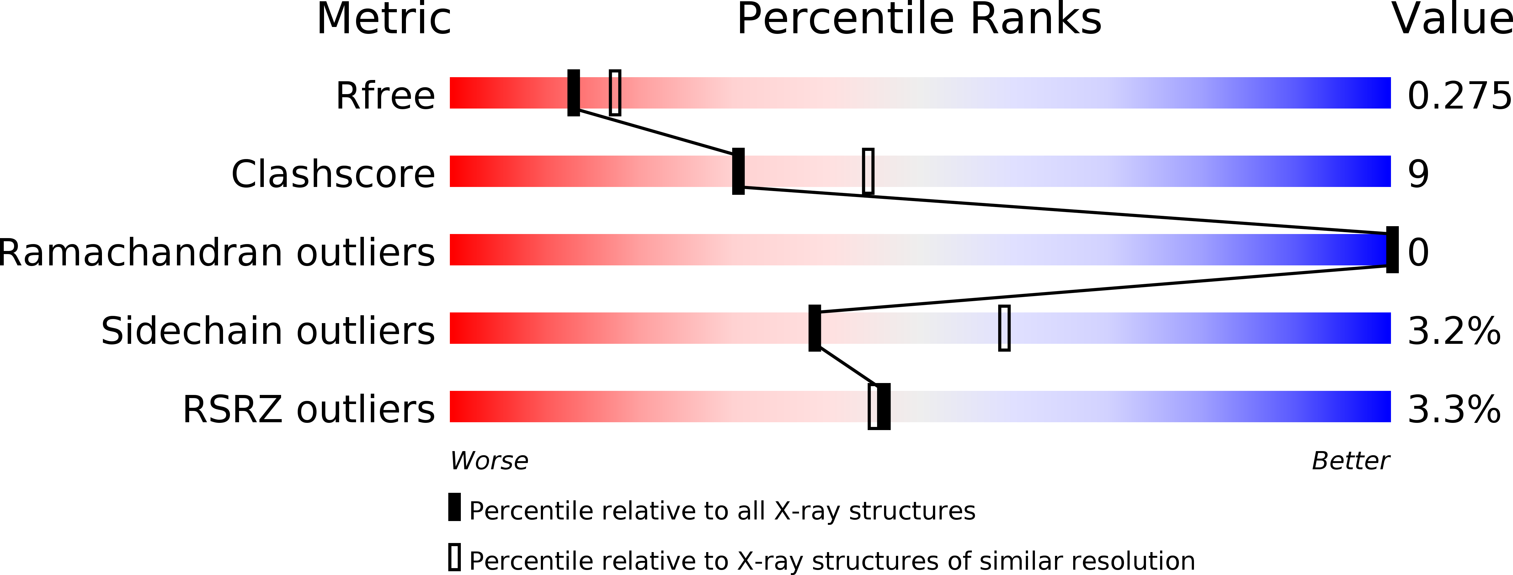
Resolution:
2.40 Å
R-Value Free:
0.27
R-Value Work:
0.19
R-Value Observed:
0.20
Space Group:
C 1 2 1
Macromolecular Entities
Structures with similar UniProt ID
Protein Blast
Polymer Type:polypeptide(L)
Molecule:Adenylation domain
Gene (Uniprot):thr1
Chain IDs:A, B
Chain Length:529
Number of Molecules:2
Biological Source:Streptomyces sp.
Primary Citation
Structure of the adenylation domain Thr1 involved in the biosynthesis of 4-chlorothreonine in Streptomyces sp. OH-5093-protein flexibility and molecular bases of substrate specificity.
FEBS J. 284 2981 2999 (2017)
PMID: 28704585 DOI: 10.1111/febs.14163

Abstact

UNLABELLED We determined the crystal structure of Thr1, the self-standing adenylation domain involved in the nonribosomal-like biosynthesis of free 4-chlorothreonine in Streptomyces sp. OH-5093. Thr1 shows two monomers in the crystallographic asymmetric unit with different relative orientations of the C- and N-terminal subdomains both in the presence of substrates and in the unliganded form. Cocrystallization with substrates, adenosine 5'-triphosphate and l-threonine, yielded one monomer containing the two substrates and the other in complex with l-threonine adenylate, locked in a postadenylation state. Steady-state kinetics showed that Thr1 activates l-Thr and its stereoisomers, as well as d-Ala, l- and d-Ser, albeit with lower efficiency. Modeling of these substrates in the active site highlighted the molecular bases of substrate discrimination. This work provides the first crystal structure of a threonine-activating adenylation enzyme, a contribution to the studies on conformational rearrangement in adenylation domains and on substrate recognition in nonribosomal biosynthesis. DATABASE Structural data are available in the Protein Data Bank under the accession number 5N9W and 5N9X.

Legend

Protein

Chemical

Disease

Primary Citation of related structures