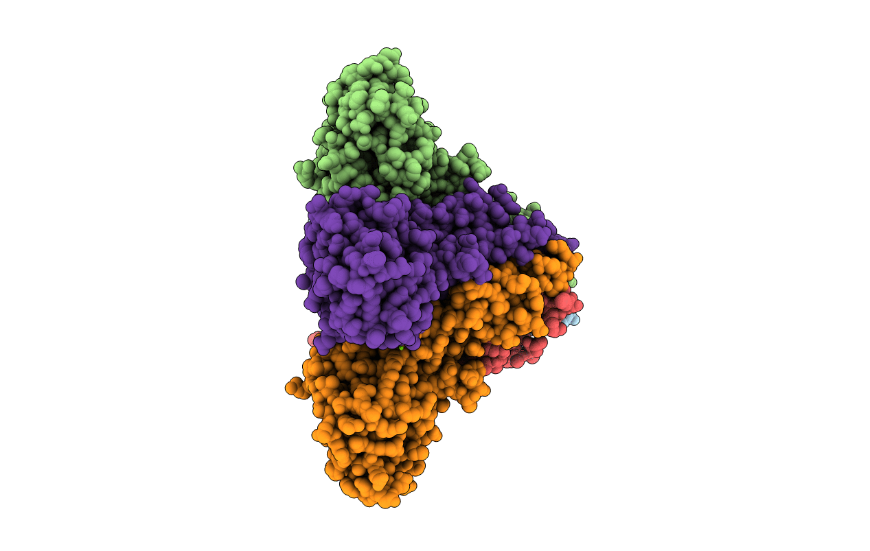
5N77 image
Entry Detail
PDB ID:
5N77
Title:
Crystal structure of the cytosolic domain of the CorA magnesium channel from Escherichia coli in complex with magnesium
Biological Source:
Source Organism:
Host Organism:
PDB Version:
Deposition Date:
2017-02-20
Release Date:
2017-07-12
Method Details:
Experimental Method:
Resolution:
2.80 Å
R-Value Free:
0.23
R-Value Work:
0.19
R-Value Observed:
0.19
Space Group:
P 1 21 1
Macromolecular Entities
Polymer Type:polypeptide(L)
Description:Magnesium transport protein CorA
Chain IDs:A, B, C, D, E
Chain Length:257
Number of Molecules:5
Biological Source:Escherichia coli
Primary Citation
Structure and Cooperativity of the Cytosolic Domain of the CorA Mg(2+) Channel from Escherichia coli.
Structure 25 1175 1186.e4 (2017)
PMID: 28669631 DOI: 10.1016/j.str.2017.05.024

Abstact

Structures of the Mg2+ bound (closed) and apo (open) states of CorA suggests that channel gating is accomplished by rigid-body motions between symmetric and asymmetric assemblies of the cytosolic portions of the five subunits in response to ligand (Mg2+) binding/unbinding at interfacial sites. Here, we structurally and biochemically characterize the isolated cytosolic domain from Escherichia coli CorA. The data reveal an Mg2+-ligand binding site located in a novel position between each of the five subunits and two Mg2+ ions trapped inside the pore. Soaking experiments show that cobalt hexammine outcompetes Mg2+ at the pore site closest to the membrane. This represents the first structural information of how an analog of hexa-hydrated Mg2+ (and competitive inhibitor of CorA) associates to the CorA pore. Biochemical data on the isolated cytoplasmic domain and full-length protein suggests that gating of the CorA channel is governed cooperatively.

Legend

Protein

Chemical

Disease

Primary Citation of related structures