5N4W image
Deposition Date 2017-02-11
Release Date 2017-06-07
Last Version Date 2024-05-01
Entry Detail
PDB ID:
5N4W
Keywords:
Title:
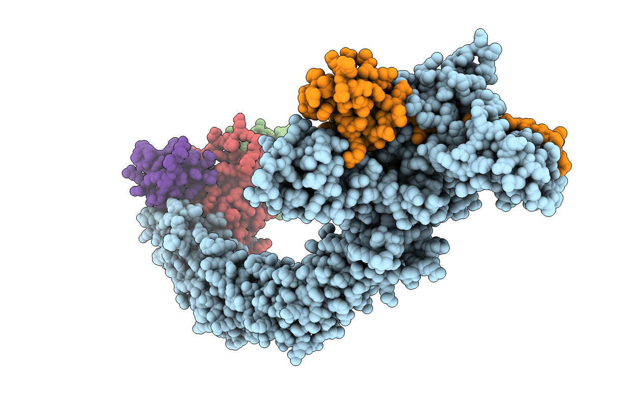
Crystal structure of the Cul2-Rbx1-EloBC-VHL ubiquitin ligase complex
Biological Source:
Source Organism(s):
Homo sapiens (Taxon ID: 9606)
Method Details:
Experimental Method:
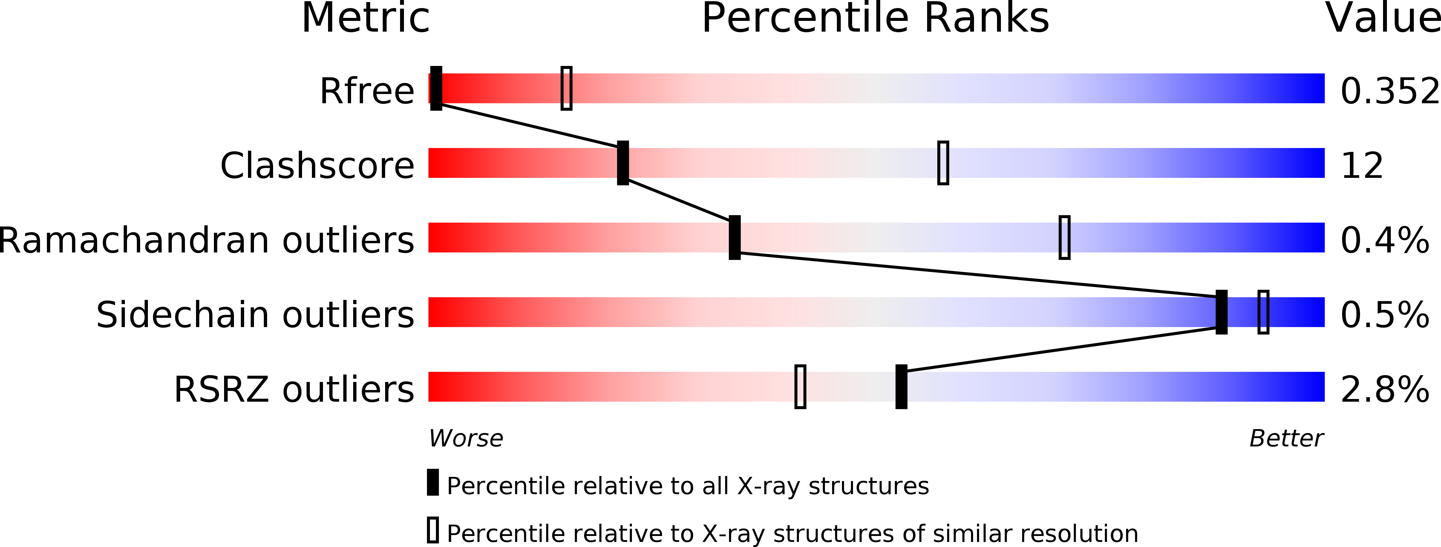
Resolution:
3.90 Å
R-Value Free:
0.34
R-Value Work:
0.30
R-Value Observed:
0.30
Space Group:
C 2 2 21
Macromolecular Entities
Structures with similar UniProt ID
Protein Blast
Polymer Type:polypeptide(L)
Molecule:Cullin-2
Gene (Uniprot):CUL2
Chain IDs:A
Chain Length:748
Number of Molecules:1
Biological Source:Homo sapiens
Structures with similar UniProt ID
Protein Blast
Polymer Type:polypeptide(L)
Molecule:Elongin-B
Gene (Uniprot):ELOB
Chain IDs:D (auth: B)
Chain Length:104
Number of Molecules:1
Biological Source:Homo sapiens
Structures with similar UniProt ID
Protein Blast
Polymer Type:polypeptide(L)
Molecule:Elongin-C
Gene (Uniprot):ELOC
Chain IDs:E (auth: C)
Chain Length:97
Number of Molecules:1
Biological Source:Homo sapiens
Structures with similar UniProt ID
Protein Blast
Polymer Type:polypeptide(L)
Molecule:E3 ubiquitin-protein ligase RBX1
Gene (Uniprot):RBX1
Chain IDs:C (auth: R)
Chain Length:102
Number of Molecules:1
Biological Source:Homo sapiens
Structures with similar UniProt ID
Protein Blast
Polymer Type:polypeptide(L)
Molecule:Von Hippel-Lindau disease tumor suppressor
Gene (Uniprot):VHL
Chain IDs:B (auth: V)
Chain Length:160
Number of Molecules:1
Biological Source:Homo sapiens
Ligand Molecules
Primary Citation
Crystal Structure of the Cul2-Rbx1-EloBC-VHL Ubiquitin Ligase Complex.
Structure 25 901 911.e3 (2017)
PMID: 28591624 DOI: 10.1016/j.str.2017.04.009

Abstact

Cullin RING E3 ubiquitin ligases (CRLs) function in the ubiquitin proteasome system to catalyze the transfer of ubiquitin from E2 conjugating enzymes to specific substrate proteins. CRLs are large dynamic complexes and attractive drug targets for the development of small-molecule inhibitors and chemical inducers of protein degradation. The atomic details of whole CRL assembly and interactions that dictate subunit specificity remain elusive. Here we present the crystal structure of a pentameric CRL2VHL complex, composed of Cul2, Rbx1, Elongin B, Elongin C, and pVHL. The structure traps a closed state of full-length Cul2 and a new pose of Rbx1 in a trajectory from closed to open conformation. We characterize hotspots and binding thermodynamics at the interface between Cul2 and pVHL-EloBC and identify mutations that contribute toward a selectivity switch for Cul2 versus Cul5 recognition. Our findings provide structural and biophysical insights into the whole Cul2 complex that could aid future drug targeting.

Legend

Protein

Chemical

Disease

Primary Citation of related structures
Feedback Form
Name
Email
Institute
Feedback