Deposition Date
2017-02-10
Release Date
2018-02-14
Last Version Date
2024-11-20
Entry Detail
PDB ID:
5N48
Keywords:
Title:
Structure of Anticalin N9B in complex with extra-domain B of human oncofetal fibronectin
Biological Source:
Source Organism(s):
Homo sapiens (Taxon ID: 9606)
Expression System(s):
Method Details:
Experimental Method:
Resolution:
1.60 Å
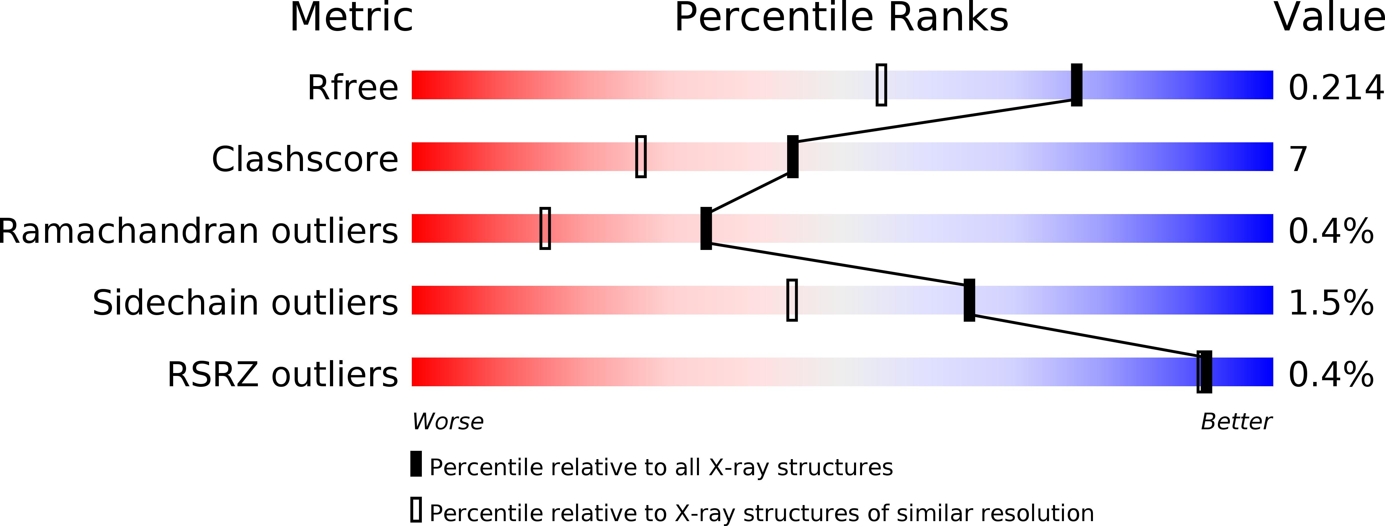
R-Value Free:
0.20
R-Value Work:
0.16
R-Value Observed:
0.17
Space Group:
P 1 21 1