5N47 image
Deposition Date 2017-02-10
Release Date 2018-02-14
Last Version Date 2024-10-23
Entry Detail
PDB ID:
5N47
Keywords:
Title:
Structure of Anticalin N7E in complex with the three-domain fragment Fn7B8 of human oncofetal fibronectin
Biological Source:
Source Organism:
Homo sapiens (Taxon ID: 9606)
Method Details:
Experimental Method:
Resolution:
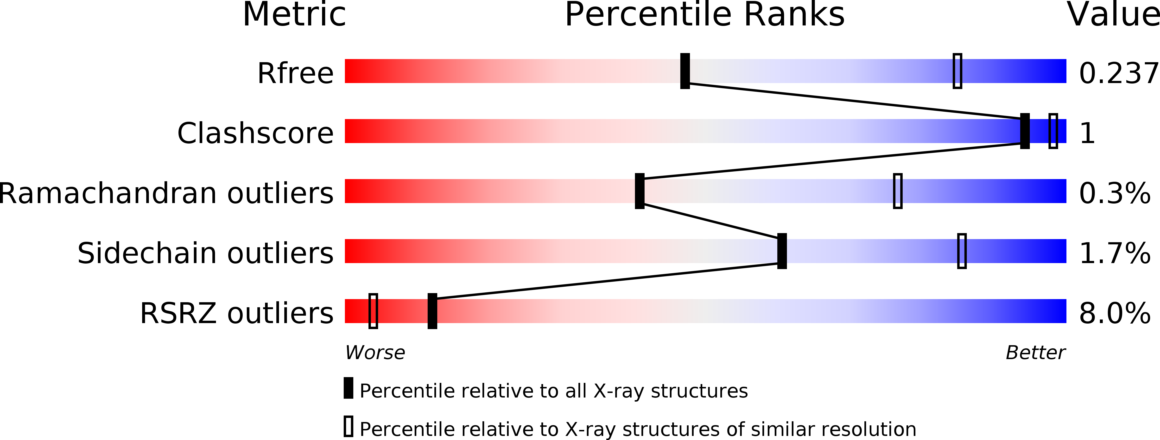
3.00 Å
R-Value Free:
0.24
R-Value Work:
0.20
R-Value Observed:
0.20
Space Group:
P 41 21 2
Macromolecular Entities
Structures with similar UniProt ID
Protein Blast
Polymer Type:polypeptide(L)
Molecule:Neutrophil gelatinase-associated lipocalin
Gene (Uniprot):LCN2
Mutagens:Q28H, L36E, A40S, I41L, Q49R, L70R, K73S, D77H, W79L, C87S, N96L, Y100K, L103H, Y106F, K125T, S127A, K134F
Chain IDs:A, C, E
Chain Length:188
Number of Molecules:3
Biological Source:Homo sapiens
Structures with similar UniProt ID
Protein Blast
Polymer Type:polypeptide(L)
Molecule:Fibronectin
Gene (Uniprot):FN1
Chain IDs:B, D, F
Chain Length:284
Number of Molecules:3
Biological Source:Homo sapiens
Ligand Molecules
Primary Citation
Anticalins Reveal High Plasticity in the Mode of Complex Formation with a Common Tumor Antigen.
Structure 26 649 656.e3 (2018)
PMID: 29526433 DOI: 10.1016/j.str.2018.02.003

Abstact

We describe the comparative X-ray structural analysis of three Anticalin proteins directed against the extra-domain B (ED-B) of oncofetal fibronectin (Fn), a validated marker of tumor neoangiogenesis. The Anticalins were engineered from the human lipocalin 2 (Lcn2) scaffold via targeted randomization of the structurally variable loop region and selection by phage display, resulting in 15-19 exchanged residues. While the four reshaped loops exhibit diverse conformations (with shifts in Cα positions up to 20.4 Å), the β-barrel core of the lipocalin remains strongly conserved, thus confirming the extraordinary robustness of this scaffold. All three Anticalins bind the cc' hairpin loop of ED-B, the most exposed motif in the context of its neighboring Fn domains, but reveal entirely different binding modes, with orientations differing by up to 180°. Hence, each Anticalin recognizes its molecular target in an individual manner, in line with the distinct epitope specificities previously seen in binding experiments.

Legend

Protein

Chemical

Disease

Primary Citation of related structures