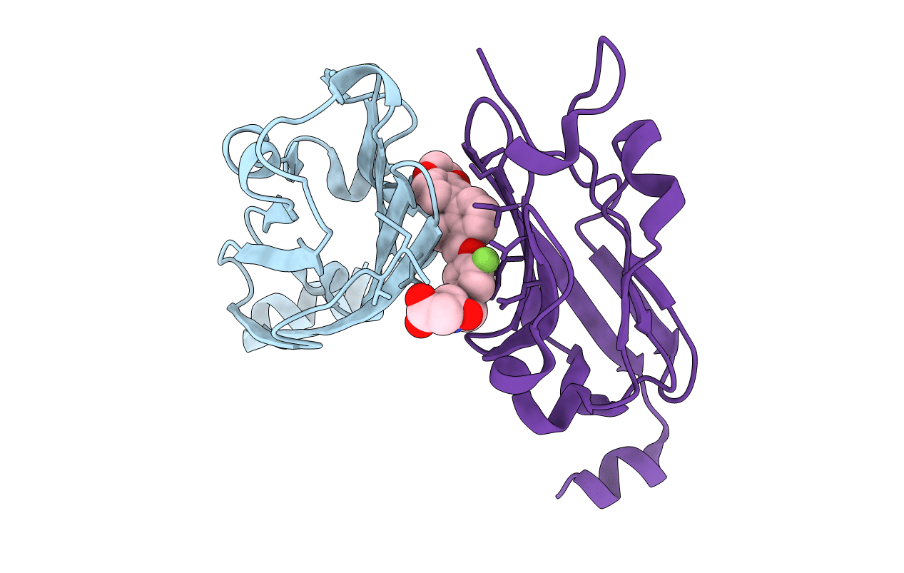
5N2F image
Deposition Date 2017-02-07
Release Date 2017-06-28
Last Version Date 2024-11-13
Entry Detail
PDB ID:
5N2F
Keywords:
Title:
Structure of PD-L1/small-molecule inhibitor complex
Biological Source:
Source Organism:
Homo sapiens (Taxon ID: 9606)
Host Organism:
Method Details:
Experimental Method:
Resolution:
1.70 Å
R-Value Free:
0.22
R-Value Work:
0.19
R-Value Observed:
0.19
Space Group:
P 2 21 21
Macromolecular Entities
Structures with similar UniProt ID
Protein Blast
Polymer Type:polypeptide(L)
Molecule:Programmed cell death 1 ligand 1
Gene (Uniprot):CD274
Chain IDs:A
Chain Length:126
Number of Molecules:1
Biological Source:Homo sapiens
Structures with similar UniProt ID
Protein Blast
Polymer Type:polypeptide(L)
Molecule:Programmed cell death 1 ligand 1
Gene (Uniprot):CD274
Chain IDs:B
Chain Length:124
Number of Molecules:1
Biological Source:Homo sapiens
Ligand Molecules
Primary Citation
Small-Molecule Inhibitors of the Programmed Cell Death-1/Programmed Death-Ligand 1 (PD-1/PD-L1) Interaction via Transiently Induced Protein States and Dimerization of PD-L1.
J. Med. Chem. 60 5857 5867 (2017)
PMID: 28613862 DOI: 10.1021/acs.jmedchem.7b00293

Abstact

Blockade of the PD-1/PD-L1 immune checkpoint pathway with monoclonal antibodies has provided significant advances in cancer treatment. The antibody-based immunotherapies carry a number of disadvantages such as the high cost of the antibodies, their limited half-life, and immunogenicity. Development of small-molecule PD-1/PD-L1 inhibitors that could overcome these drawbacks is slow because of the incomplete structural information for this pathway. The first chemical PD-1/PD-L1 inhibitors have been recently disclosed by Bristol-Myers Squibb. Here we present NMR and X-ray characterization for the two classes of these inhibitors. The X-ray structures of the PD-L1/inhibitor complexes reveal one inhibitor molecule located at the center of the PD-L1 homodimer, filling a deep hydrophobic channel-like pocket between two PD-L1 molecules. Derivatives of (2-methyl-3-biphenylyl)methanol exhibit the structures capped on one side of the channel, whereas the compounds based on [3-(2,3-dihydro-1,4-benzodioxin-6-yl)-2-methylphenyl]methanol induce an enlarged interaction interface that results in the open "face-back" tunnel through the PD-L1 dimer.

Legend

Protein

Chemical

Disease

Primary Citation of related structures