5N1T image
Deposition Date 2017-02-06
Release Date 2018-02-28
Last Version Date 2025-10-01
Entry Detail
PDB ID:
5N1T
Keywords:
Title:
Crystal structure of complex between flavocytochrome c and copper chaperone CopC from T. paradoxus
Biological Source:
Source Organism:
Method Details:
Experimental Method:
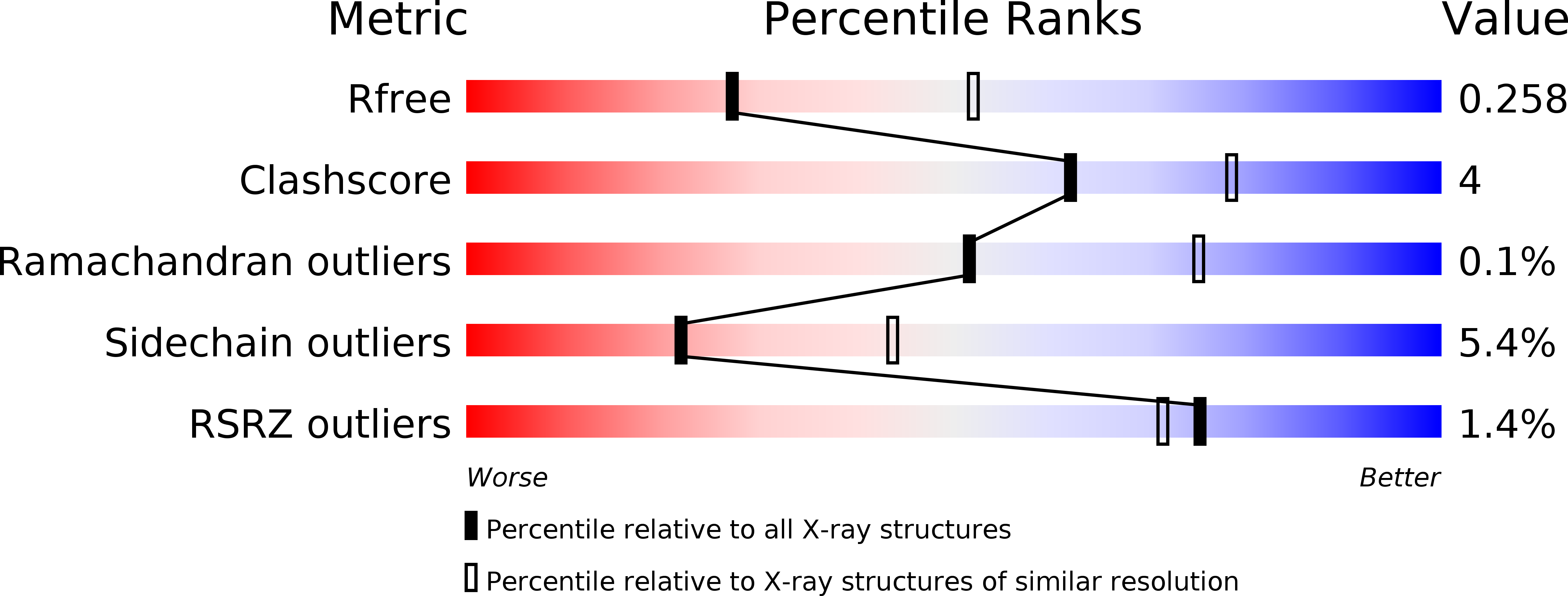
Resolution:
2.60 Å
R-Value Free:
0.25
R-Value Work:
0.18
R-Value Observed:
0.18
Space Group:
P 21 21 21
Macromolecular Entities
Structures with similar UniProt ID
Protein Blast
Polymer Type:polypeptide(L)
Molecule:flavin-binding subunit of sulfide dehydrogenase
Gene (Uniprot):THITH_13285
Mutagens:C199CSS
Chain IDs:A
Chain Length:429
Number of Molecules:1
Biological Source:Thioalkalivibrio paradoxus ARh 1
Structures with similar UniProt ID
Protein Blast
Polymer Type:polypeptide(L)
Molecule:Cytochrome C
Gene (Uniprot):THITH_13280
Chain IDs:B
Chain Length:102
Number of Molecules:1
Biological Source:Thioalkalivibrio paradoxus ARh 1
Structures with similar UniProt ID
Protein Blast
Polymer Type:polypeptide(L)
Molecule:CopC
Gene (Uniprot):THITH_13340
Chain IDs:C (auth: M), D (auth: W)
Chain Length:160
Number of Molecules:2
Biological Source:Thioalkalivibrio paradoxus ARh 1
Modified Residue
Compound ID Chain ID Parent Comp ID Details 2D Image
CSS A CYS modified residue
Primary Citation
Structure of the flavocytochrome c sulfide dehydrogenase associated with the copper-binding protein CopC from the haloalkaliphilic sulfur-oxidizing bacterium Thioalkalivibrio paradoxusARh 1.
Acta Crystallogr D Struct Biol 74 632 642 (2018)
PMID: 29968673 DOI: 10.1107/S2059798318005648

Abstact

Flavocytochrome c sulfide dehydrogenase from Thioalkalivibrio paradoxus (TpFCC) is a heterodimeric protein consisting of flavin- and monohaem c-binding subunits. TpFCC was co-purified and co-crystallized with the dimeric copper-binding protein TpCopC. The structure of the TpFCC-(TpCopC)2 complex was determined by X-ray diffraction at 2.6 Å resolution. The flavin-binding subunit of TpFCC is structurally similar to those determined previously, and the structure of the haem-binding subunit is similar to that of the N-terminal domain of dihaem FCCs. According to classification based on amino-acid sequence, TpCopC belongs to a high-affinity CopC subfamily characterized by the presence of a conserved His1-Xxx-His3 motif at the N-terminus. Apparently, a unique α-helix which is present in each monomer of TpCopC at the interface with TpFCC plays a key role in complex formation. The structure of the copper-binding site in TpCopC is similar to those in other known CopC structures. His3 is not involved in binding to the copper ion and is 6-7 Å away from this ion. Therefore, the His1-Xxx-His3 motif cannot be considered to be a key factor in the high affinity of CopC for copper(II) ions. It is suggested that the TpFCC-(TpCopC)2 heterotetramer may be a component of a large periplasmic complex that is responsible for thiocyanate metabolism.

Legend

Protein

Chemical

Disease

Primary Citation of related structures
Feedback Form
Name
Email
Institute
Feedback