5MXK image
Deposition Date 2017-01-23
Release Date 2017-05-31
Last Version Date 2024-01-17
Entry Detail
PDB ID:
5MXK
Keywords:
Title:
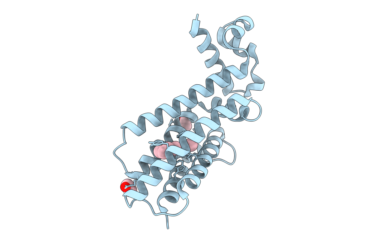
Structure of Mycobacterium Tuberculosis Transcriptional Regulatory Repressor Protein (EthR) in complex with fragment 7G9.
Biological Source:
Source Organism:
Method Details:
Experimental Method:
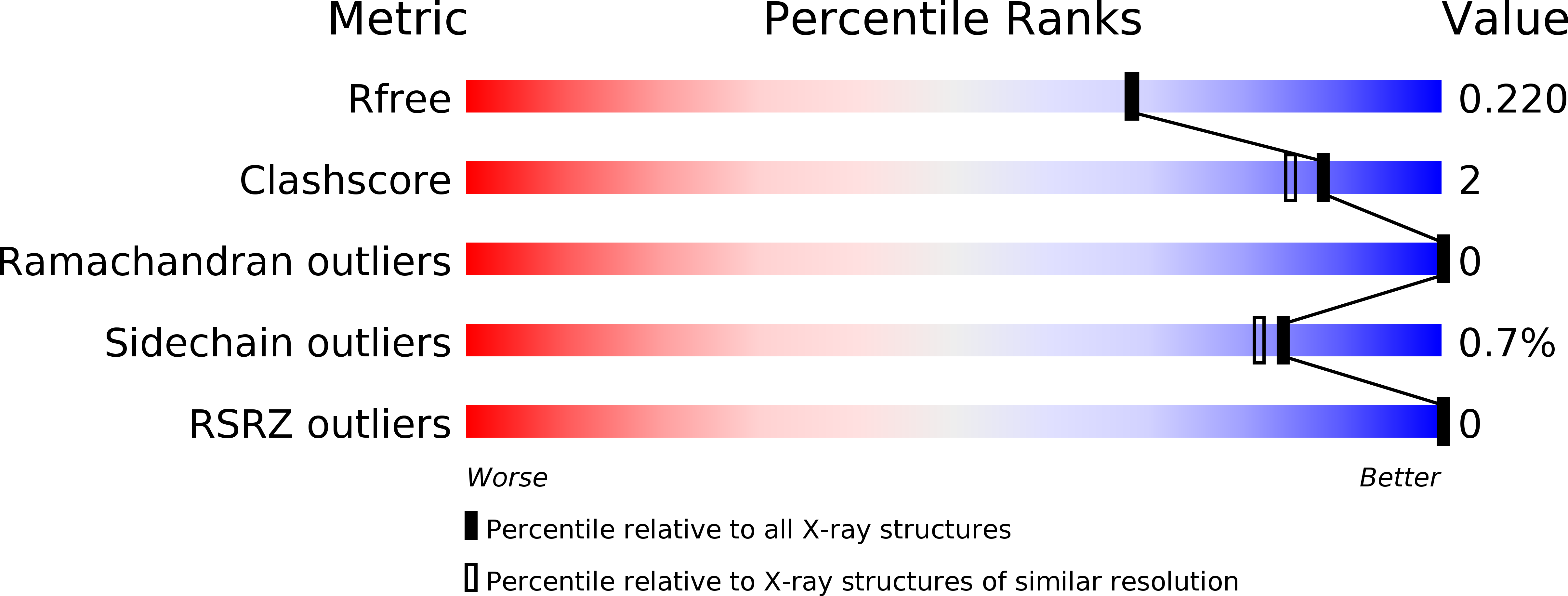
Resolution:
1.93 Å
R-Value Free:
0.22
R-Value Work:
0.18
R-Value Observed:
0.18
Space Group:
P 41 21 2
Macromolecular Entities
Structures with similar UniProt ID
Protein Blast
Polymer Type:polypeptide(L)
Molecule:HTH-type transcriptional regulator EthR
Gene (Uniprot):ethR
Chain IDs:A
Chain Length:216
Number of Molecules:1
Biological Source:Mycobacterium tuberculosis H37Rv
Primary Citation
Fragment Screening against the EthR-DNA Interaction by Native Mass Spectrometry.
Angew. Chem. Int. Ed. Engl. 56 7488 7491 (2017)
PMID: 28513917 DOI: 10.1002/anie.201702888

Abstact

Native nanoelectrospray ionization mass spectrometry is an underutilized technique for fragment screening. In this study, the first demonstration is provided of the use of native mass spectrometry for screening fragments against a protein-DNA interaction. EthR is a transcriptional repressor of EthA expression in Mycobacterium tuberculosis (Mtb) that reduces the efficacy of ethionamide, a second-line antitubercular drug used to combat multidrug-resistant Mtb strains. A small-scale fragment screening campaign was conducted against the EthR-DNA interaction using native mass spectrometry, and the results were compared with those from differential scanning fluorimetry, a commonly used primary screening technique. Hits were validated by surface plasmon resonance and X-ray crystallography. The screening campaign identified two new fragments that disrupt the EthR-DNA interaction in vitro (IC50 =460-610 μm) and bind to the hydrophobic channel of the EthR dimer.

Legend

Protein

Chemical

Disease

Primary Citation of related structures
Feedback Form
Name
Email
Institute
Feedback