5MX0 image
Deposition Date 2017-01-20
Release Date 2017-03-01
Last Version Date 2024-11-13
Entry Detail
PDB ID:
5MX0
Title:
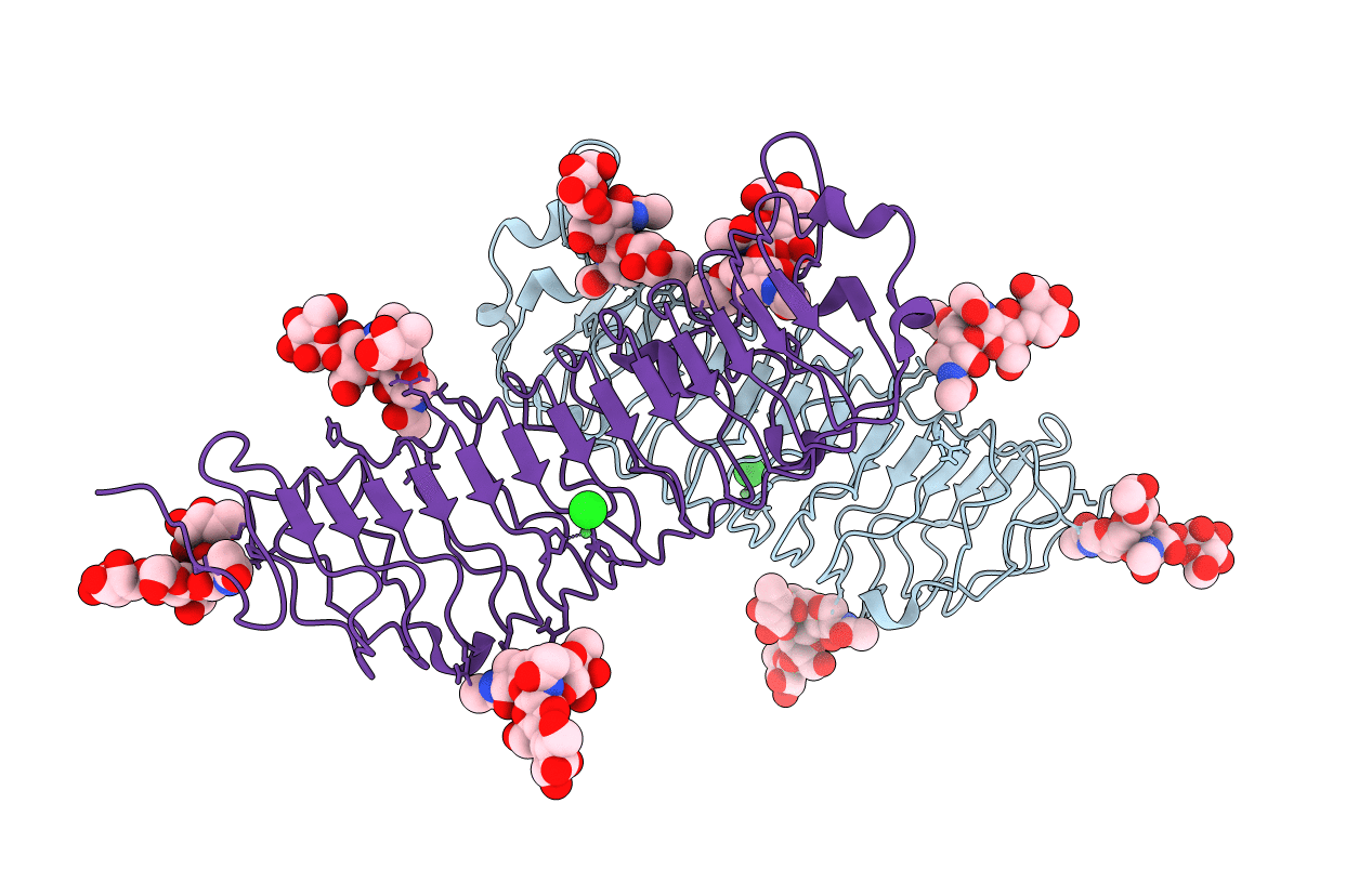
Crystal structure of human fibromodulin
Biological Source:
Source Organism:
Homo sapiens (Taxon ID: 9606)
Host Organism:
Method Details:
Experimental Method:
Resolution:
2.21 Å
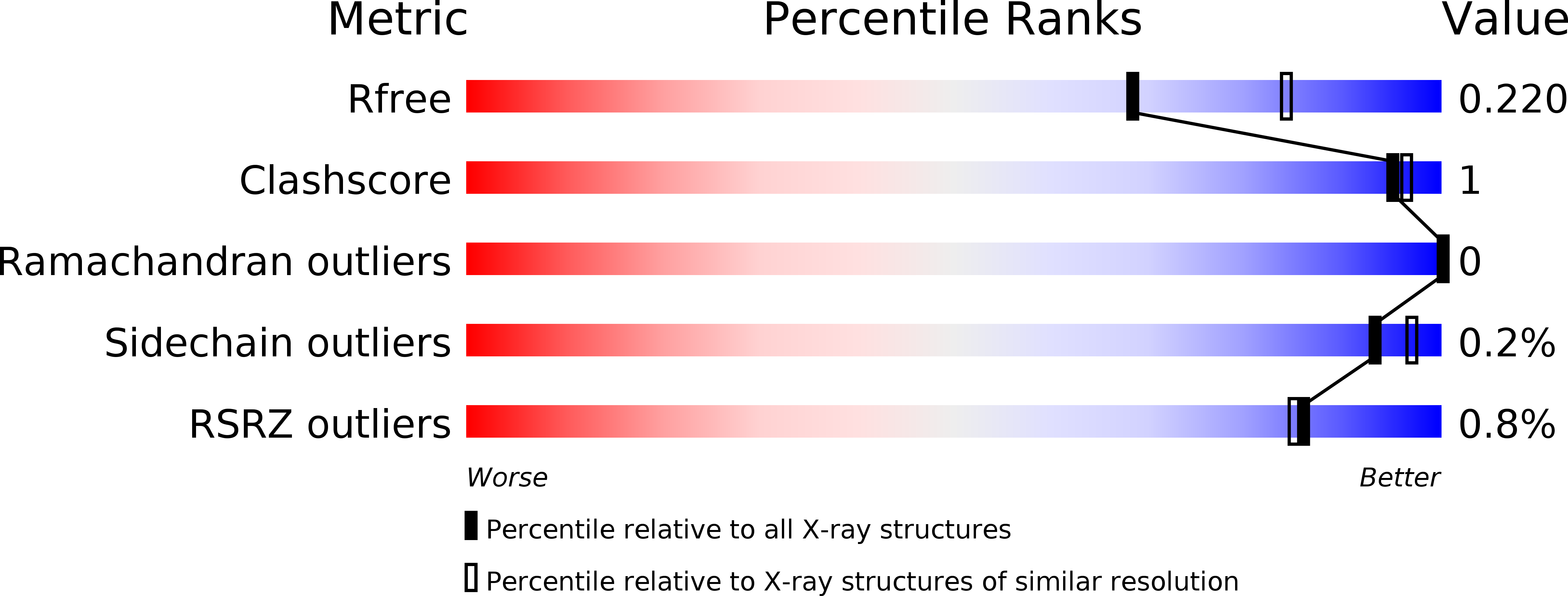
R-Value Free:
0.21
R-Value Work:
0.17
R-Value Observed:
0.18
Space Group:
C 1 2 1
Macromolecular Entities
Polymer Type:polypeptide(L)
Molecule:Fibromodulin
Gene (Uniprot):FMOD
Mutations:Y38S, Y39S, Y42S, Y45S, Y47S, Y50S, Y53S, Y55S, Y63S, Y65S
Chain IDs:A, B
Chain Length:362
Number of Molecules:2
Biological Source:Homo sapiens
Primary Citation
Structural and functional analysis of two small leucine-rich repeat proteoglycans, fibromodulin and chondroadherin.
Matrix Biol. 63 106 116 (2017)
PMID: 28215822 DOI: 10.1016/j.matbio.2017.02.002

Abstact

The small leucine-rich proteoglycans (SLRPs) are important regulators of extracellular matrix assembly and cell signalling. We have determined crystal structures at ~2.2Å resolution of human fibromodulin and chondroadherin, two collagen-binding SLRPs. Their overall fold is similar to that of the prototypical SLRP, decorin, but unlike decorin neither fibromodulin nor chondroadherin forms a stable dimer. A previously identified binding site for integrin α2β1 maps to an α-helix in the C-terminal cap region of chondroadherin. Interrogation of the Collagen Toolkits revealed a unique binding site for chondroadherin in collagen II, and no binding to collagen III. A triple-helical peptide containing the sequence GAOGPSGFQGLOGPOGPO (O is hydroxyproline) forms a stable complex with chondroadherin in solution. In fibrillar collagen I and II, this sequence is aligned with the collagen cross-linking site KGHR, suggesting a role for chondroadherin in cross-linking.

Legend

Protein

Chemical

Disease

Primary Citation of related structures