5MWN image
Deposition Date 2017-01-18
Release Date 2017-06-28
Last Version Date 2024-10-16
Entry Detail
PDB ID:
5MWN
Title:
Structure of the EAEC T6SS component TssK N-terminal domain in complex with llama nanobodies nbK18 and nbK27
Biological Source:
Source Organism:
Escherichia coli (Taxon ID: 562)
Lama glama (Taxon ID: 9844)
Host Organism:
Method Details:
Experimental Method:
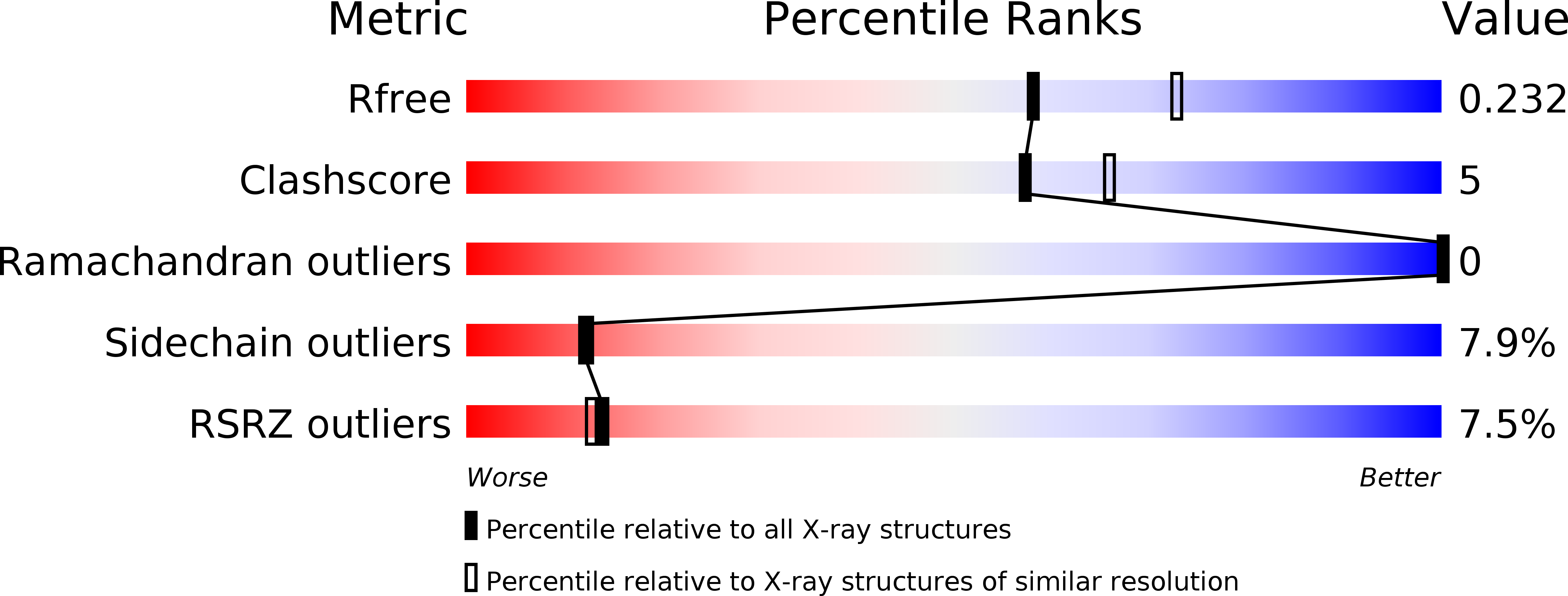
Resolution:
2.20 Å
R-Value Free:
0.22
R-Value Work:
0.19
R-Value Observed:
0.19
Space Group:
P 21 21 21
Macromolecular Entities
Polymer Type:polypeptide(L)
Molecule:Type VI secretion protein
Chain IDs:A, B, C
Chain Length:315
Number of Molecules:3
Biological Source:Escherichia coli
Polymer Type:polypeptide(L)
Molecule:llama nanobody raised against TssK, nbK18
Chain IDs:D, E, F
Chain Length:125
Number of Molecules:3
Biological Source:Lama glama
Polymer Type:polypeptide(L)
Molecule:llama nanobody raised against TssK, nbK27
Chain IDs:G (auth: N)
Chain Length:125
Number of Molecules:1
Biological Source:Lama glama
Ligand Molecules
Primary Citation
Type VI secretion TssK baseplate protein exhibits structural similarity with phage receptor-binding proteins and evolved to bind the membrane complex.
Nat Microbiol 2 17103 17103 (2017)
PMID: 28650463 DOI: 10.1038/nmicrobiol.2017.103

Abstact

The type VI secretion system (T6SS) is a multiprotein machine widespread in Gram-negative bacteria that delivers toxins into both eukaryotic and prokaryotic cells. The mechanism of action of the T6SS is comparable to that of contractile myophages. The T6SS builds a tail-like structure made of an inner tube wrapped by a sheath, assembled under an extended conformation. Contraction of the sheath propels the inner tube towards the target cell. The T6SS tail is assembled on a platform-the baseplate-which is functionally similar to bacteriophage baseplates. In addition, the baseplate docks the tail to a trans-envelope membrane complex that orients the tail towards the target. Here, we report the crystal structure of TssK, a central component of the T6SS baseplate. We show that TssK is composed of three domains, and establish the contribution of each domain to the interaction with TssK partners. Importantly, this study reveals that the N-terminal domain of TssK is structurally homologous to the shoulder domain of phage receptor-binding proteins, and the C-terminal domain binds the membrane complex. We propose that TssK has conserved the domain of attachment to the virion particle but has evolved the reception domain to use the T6SS membrane complex as receptor.

Legend

Protein

Chemical

Disease

Primary Citation of related structures