5MVF image
Deposition Date 2017-01-16
Release Date 2017-03-08
Last Version Date 2024-01-17
Entry Detail
PDB ID:
5MVF
Keywords:
Title:
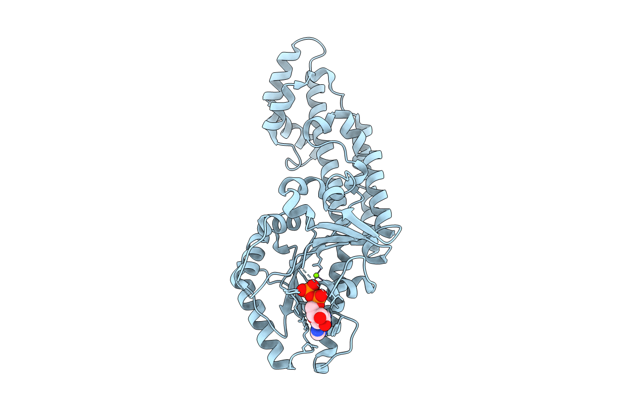
Active structure of EHD4 complexed with ADP
Biological Source:
Source Organism:
Mus musculus (Taxon ID: 10090)
Host Organism:
Method Details:
Experimental Method:
Resolution:
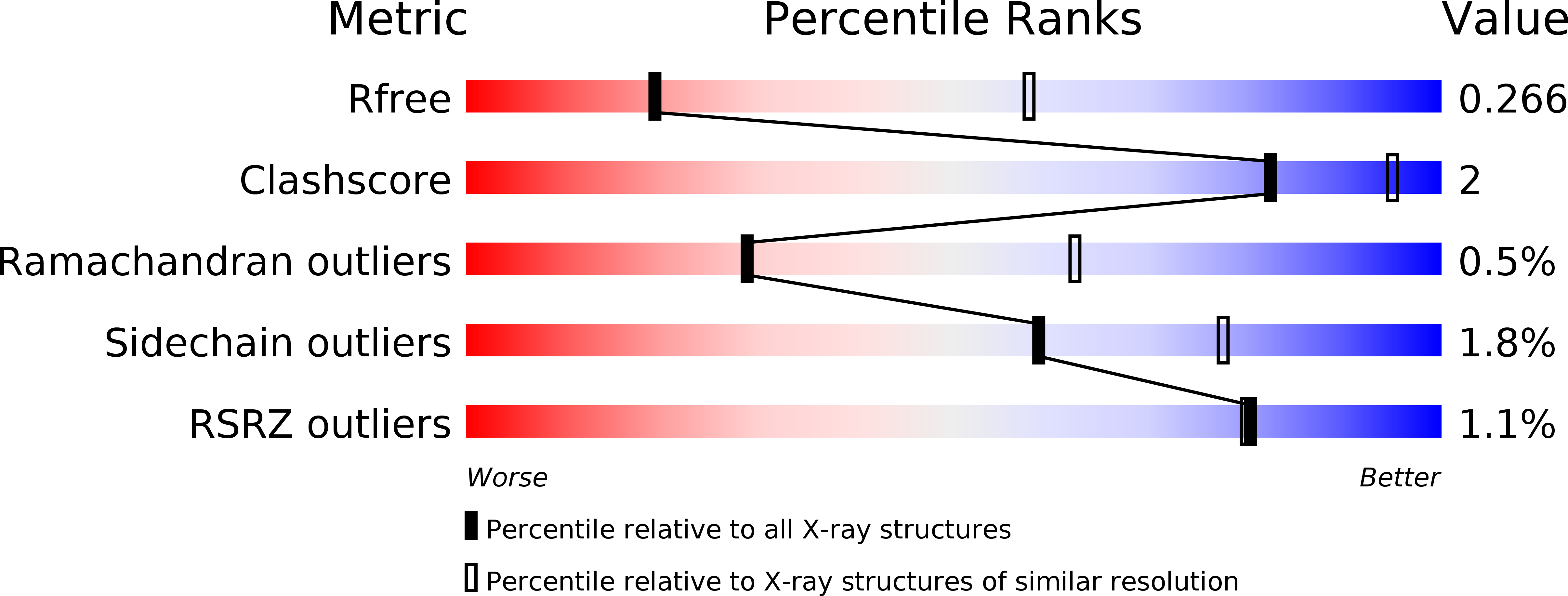
3.27 Å
R-Value Free:
0.26
R-Value Work:
0.23
R-Value Observed:
0.23
Space Group:
P 42 21 2
Macromolecular Entities
Polymer Type:polypeptide(L)
Molecule:EH domain-containing protein 4
Gene (Uniprot):Ehd4
Chain IDs:A
Chain Length:523
Number of Molecules:1
Biological Source:Mus musculus
Primary Citation
Structural insights into the activation mechanism of dynamin-like EHD ATPases.
Proc. Natl. Acad. Sci. U.S.A. 114 5629 5634 (2017)
PMID: 28228524 DOI: 10.1073/pnas.1614075114

Abstact

Eps15 (epidermal growth factor receptor pathway substrate 15)-homology domain containing proteins (EHDs) comprise a family of dynamin-related mechano-chemical ATPases involved in cellular membrane trafficking. Previous studies have revealed the structure of the EHD2 dimer, but the molecular mechanisms of membrane recruitment and assembly have remained obscure. Here, we determined the crystal structure of an amino-terminally truncated EHD4 dimer. Compared with the EHD2 structure, the helical domains are 50° rotated relative to the GTPase domain. Using electron paramagnetic spin resonance (EPR), we show that this rotation aligns the two membrane-binding regions in the helical domain toward the lipid bilayer, allowing membrane interaction. A loop rearrangement in GTPase domain creates a new interface for oligomer formation. Our results suggest that the EHD4 structure represents the active EHD conformation, whereas the EHD2 structure is autoinhibited, and reveal a complex series of domain rearrangements accompanying activation. A comparison with other peripheral membrane proteins elucidates common and specific features of this activation mechanism.

Legend

Protein

Chemical

Disease

Primary Citation of related structures