5MV5 image
Deposition Date 2017-01-15
Release Date 2017-04-05
Last Version Date 2024-05-15
Entry Detail
PDB ID:
5MV5
Keywords:
Title:
Structure of deformed wing virus, a honeybee pathogen
Biological Source:
Source Organism:
Method Details:
Experimental Method:
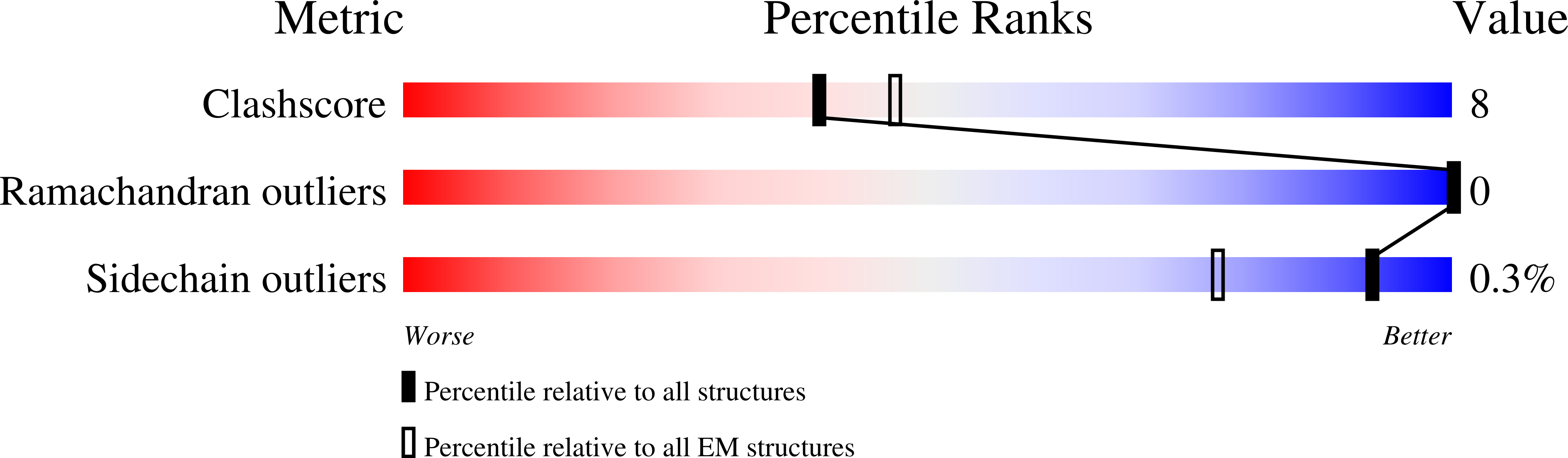
Resolution:
3.10 Å
Aggregation State:
PARTICLE
Reconstruction Method:
SINGLE PARTICLE
Macromolecular Entities
Polymer Type:polypeptide(L)
Molecule:VP1
Chain IDs:A
Chain Length:258
Number of Molecules:1
Biological Source:Deformed wing virus
Polymer Type:polypeptide(L)
Molecule:VP2
Chain IDs:B
Chain Length:253
Number of Molecules:1
Biological Source:Deformed wing virus
Polymer Type:polypeptide(L)
Molecule:VP3
Chain IDs:C
Chain Length:416
Number of Molecules:1
Biological Source:Deformed wing virus
Primary Citation
Structure of deformed wing virus, a major honey bee pathogen.
Proc. Natl. Acad. Sci. U.S.A. 114 3210 3215 (2017)
PMID: 28270616 DOI: 10.1073/pnas.1615695114

Abstact

The worldwide population of western honey bees (Apis mellifera) is under pressure from habitat loss, environmental stress, and pathogens, particularly viruses that cause lethal epidemics. Deformed wing virus (DWV) from the family Iflaviridae, together with its vector, the mite Varroa destructor, is likely the major threat to the world's honey bees. However, lack of knowledge of the atomic structures of iflaviruses has hindered the development of effective treatments against them. Here, we present the virion structures of DWV determined to a resolution of 3.1 Å using cryo-electron microscopy and 3.8 Å by X-ray crystallography. The C-terminal extension of capsid protein VP3 folds into a globular protruding (P) domain, exposed on the virion surface. The P domain contains an Asp-His-Ser catalytic triad that is, together with five residues that are spatially close, conserved among iflaviruses. These residues may participate in receptor binding or provide the protease, lipase, or esterase activity required for entry of the virus into a host cell. Furthermore, nucleotides of the DWV RNA genome interact with VP3 subunits. The capsid protein residues involved in the RNA binding are conserved among honey bee iflaviruses, suggesting a putative role of the genome in stabilizing the virion or facilitating capsid assembly. Identifying the RNA-binding and putative catalytic sites within the DWV virion structure enables future analyses of how DWV and other iflaviruses infect insect cells and also opens up possibilities for the development of antiviral treatments.

Legend

Protein

Chemical

Disease

Primary Citation of related structures