Deposition Date
2017-01-09
Release Date
2017-09-20
Last Version Date
2024-01-17
Entry Detail
PDB ID:
5MTC
Keywords:
Title:
Crystal structure of PDF from the Vibrio parahaemolyticus bacteriophage VP16T - crystal form I
Biological Source:
Source Organism(s):
Vibrio phage VP16T (Taxon ID: 238892)
Expression System(s):
Method Details:
Experimental Method:
Resolution:
1.70 Å
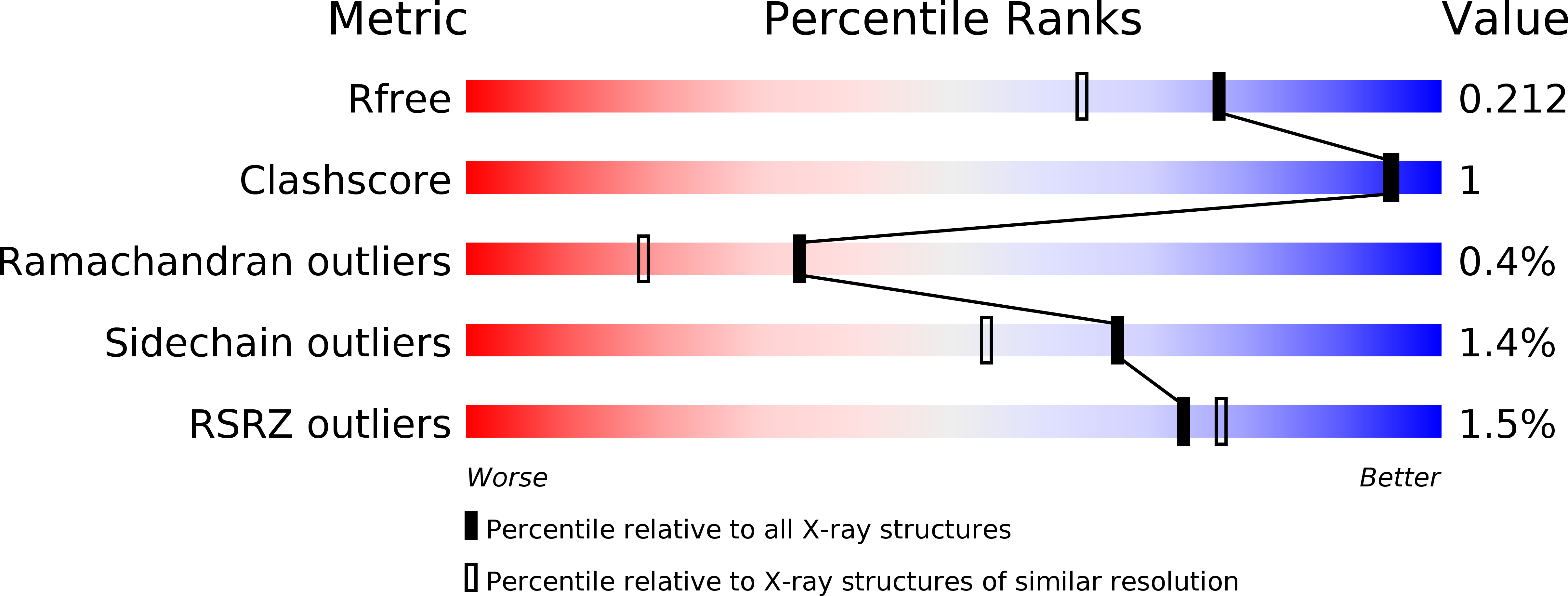
R-Value Free:
0.20
R-Value Work:
0.17
R-Value Observed:
0.17
Space Group:
P 32 2 1