
Deposition Date
2016-12-21
Release Date
2017-06-14
Last Version Date
2024-10-16
Entry Detail
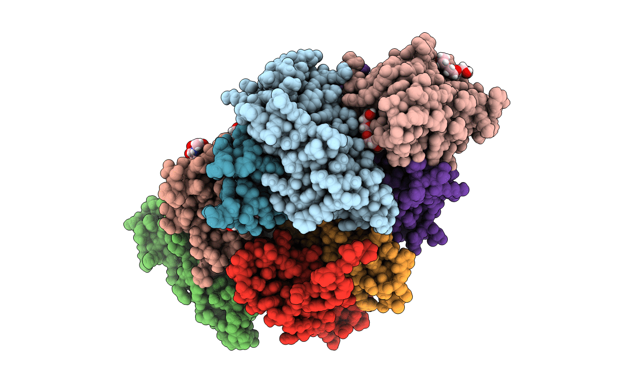
PDB ID:
5MR3
Keywords:
Title:
Crystal structure of red abalone egg VERL repeat 2 with linker in complex with sperm lysin at 1.8 A resolution
Biological Source:
Source Organism(s):
Haliotis rufescens (Taxon ID: 6454)
Expression System(s):
Method Details:
Experimental Method:
Resolution:
1.80 Å
R-Value Free:
0.22
R-Value Work:
0.20
R-Value Observed:
0.20
Space Group:
P 1 21 1
