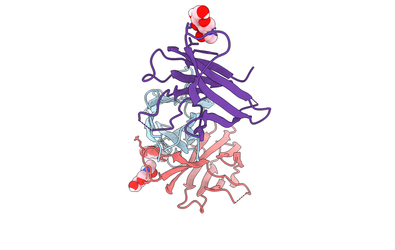
5MR2 image
Entry Detail
PDB ID:
5MR2
Keywords:
Title:
Crystal structure of red abalone VERL repeat 2 with linker at 2.5 A resolution
Biological Source:
Source Organism:
Host Organism:
PDB Version:
Deposition Date:
2016-12-21
Release Date:
2017-06-14
Method Details:
Experimental Method:
Resolution:
2.50 Å
R-Value Free:
0.28
R-Value Work:
0.23
R-Value Observed:
0.23
Space Group:
C 1 2 1
Macromolecular Entities
Polymer Type:polypeptide(L)
Description:Vitelline envelope sperm lysin receptor
Mutations:S293A, S296A, S297A
Chain IDs:A, B, C
Chain Length:134
Number of Molecules:3
Biological Source:Haliotis rufescens
Ligand Molecules
Primary Citation

Abstact

Recognition between sperm and the egg surface marks the beginning of life in all sexually reproducing organisms. This fundamental biological event depends on the species-specific interaction between rapidly evolving counterpart molecules on the gametes. We report biochemical, crystallographic, and mutational studies of domain repeats 1-3 of invertebrate egg coat protein VERL and their interaction with cognate sperm protein lysin. VERL repeats fold like the functionally essential N-terminal repeat of mammalian sperm receptor ZP2, whose structure is also described here. Whereas sequence-divergent repeat 1 does not bind lysin, repeat 3 binds it non-species specifically via a high-affinity, largely hydrophobic interface. Due to its intermediate binding affinity, repeat 2 selectively interacts with lysin from the same species. Exposure of a highly positively charged surface of VERL-bound lysin suggests that complex formation both disrupts the organization of egg coat filaments and triggers their electrostatic repulsion, thereby opening a hole for sperm penetration and fusion.

Legend

Protein

Chemical

Disease

Primary Citation of related structures