5MOY image
Entry Detail
PDB ID:
5MOY
Keywords:
Title:
Crystal structure of the BoNT/A2 receptor-binding domain in complex with the luminal domain of its neuronal receptor SV2C
Biological Source:
Host Organism:
PDB Version:
Deposition Date:
2016-12-14
Release Date:
2017-03-15
Method Details:
Experimental Method:
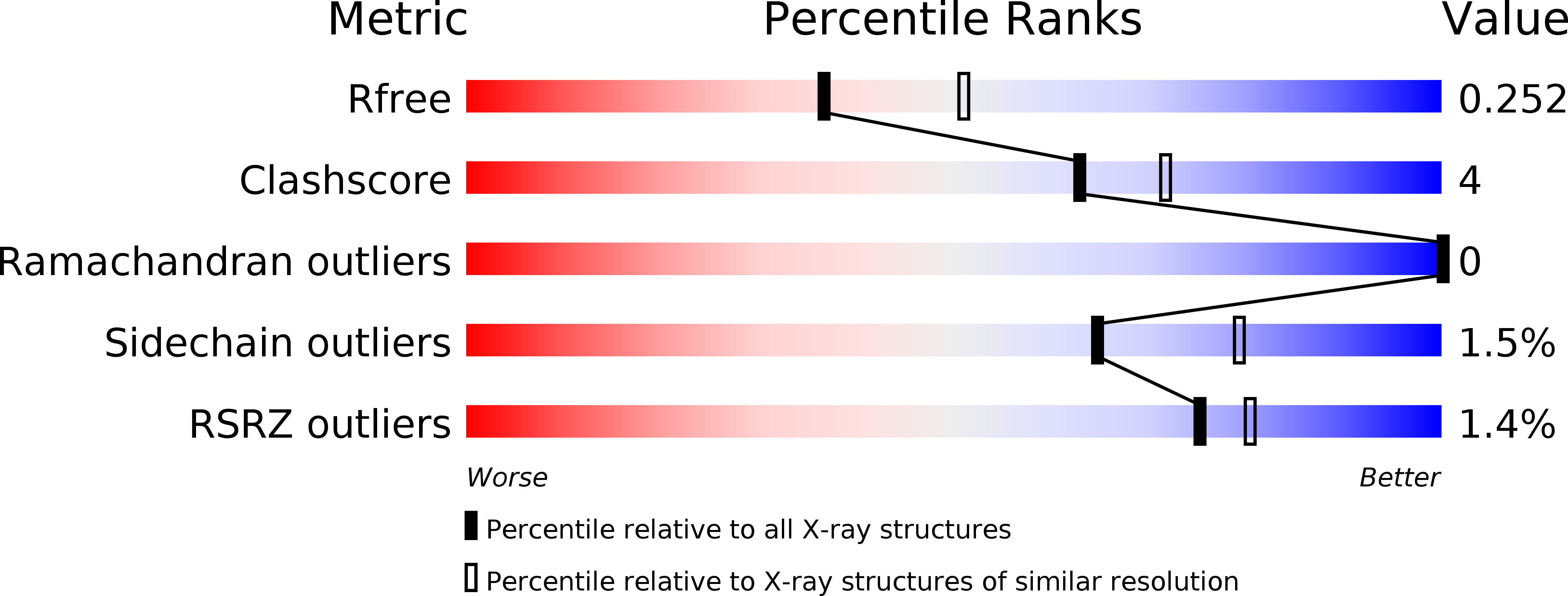
Resolution:
2.30 Å
R-Value Free:
0.25
R-Value Work:
0.19
R-Value Observed:
0.19
Space Group:
P 21 21 21
Macromolecular Entities
Polymer Type:polypeptide(L)
Description:Botulinum neurotoxin type A
Chain IDs:A
Chain Length:428
Number of Molecules:1
Biological Source:Clostridium botulinum
Polymer Type:polypeptide(L)
Description:Synaptic vesicle glycoprotein 2C
Chain IDs:B
Chain Length:136
Number of Molecules:1
Biological Source:Homo sapiens
Primary Citation
Crystal structure of the BoNT/A2 receptor-binding domain in complex with the luminal domain of its neuronal receptor SV2C.
Sci Rep 7 43588 43588 (2017)
PMID: 28252640 DOI: 10.1038/srep43588

Abstact

A detailed molecular understanding of botulinum neurotoxin (BoNT)/host-cell-receptor interactions is fundamental both for developing strategies against botulism and for generating improved BoNT variants for medical applications. The X-ray crystal structure of the receptor-binding domain (HC) of BoNT/A1 in complex with the luminal domain (LD) of its neuronal receptor SV2C revealed only few specific side-chain - side-chain interactions that are important for binding. Notably, two BoNT/A1 residues, Arg 1156 and Arg 1294, that are crucial for the interaction with SV2, are not conserved among subtypes. Because it has been suggested that differential receptor binding of subtypes might explain their differences in biological activity, we determined the crystal structure of BoNT/A2-HC in complex with SV2C-LD. Although only few side-chain interactions are conserved between the two BoNT/A subtypes, the overall binding mode of subtypes A1 and A2 is virtually identical. In the BoNT/A2-HC - SV2C complex structure, a missing cation-π stacking is compensated for by an additional salt bridge and an anion-π stacking interaction, which explains why the binding of BoNT/A subtypes to SV2C tolerates variable side chains. These findings suggest that motif extensions and a shallow binding cleft in BoNT/A-HC contribute to binding specificity.

Legend

Protein

Chemical

Disease

Primary Citation of related structures