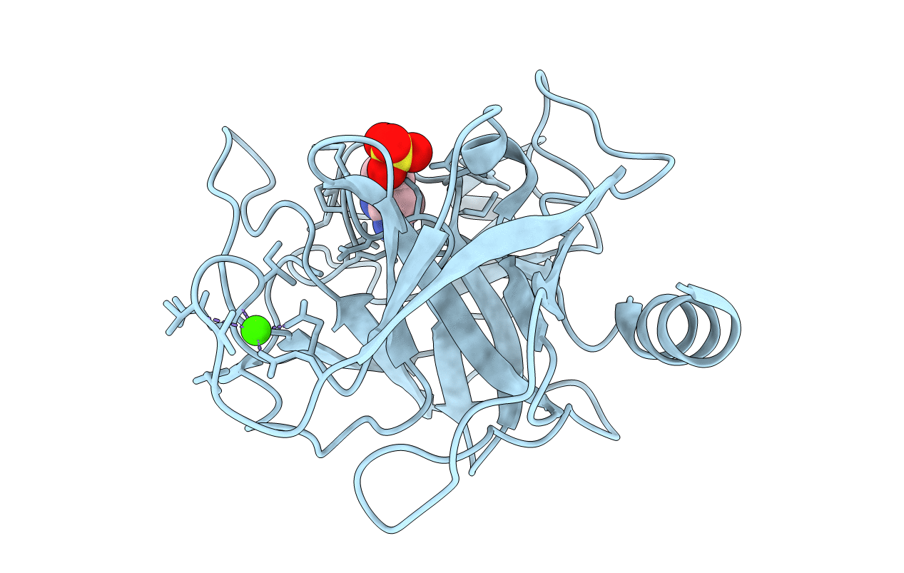
5MOS image
Entry Detail
PDB ID:
5MOS
Keywords:
Title:
Joint X-ray/neutron structure of cationic trypsin in complex with N-amidinopiperidine
Biological Source:
Source Organism:
PDB Version:
Deposition Date:
2016-12-14
Release Date:
2018-02-28
Method Details:
R-Value Free:
['0.10
R-Value Work:
['0.09
R-Value Observed:
['0.09
Space Group:
P 21 21 21
Macromolecular Entities
Polymer Type:polypeptide(L)
Description:Cationic trypsin
Chain IDs:A
Chain Length:223
Number of Molecules:1
Biological Source:Bos taurus
Primary Citation
Intriguing role of water in protein-ligand binding studied by neutron crystallography on trypsin complexes.
Nat Commun 9 3559 3559 (2018)
PMID: 30177695 DOI: 10.1038/s41467-018-05769-2

Abstact

Hydrogen bonds are key interactions determining protein-ligand binding affinity and therefore fundamental to any biological process. Unfortunately, explicit structural information about hydrogen positions and thus H-bonds in protein-ligand complexes is extremely rare and similarly the important role of water during binding remains poorly understood. Here, we report on neutron structures of trypsin determined at very high resolutions ≤1.5 Å in uncomplexed and inhibited state complemented by X-ray and thermodynamic data and computer simulations. Our structures show the precise geometry of H-bonds between protein and the inhibitors N-amidinopiperidine and benzamidine along with the dynamics of the residual solvation pattern. Prior to binding, the ligand-free binding pocket is occupied by water molecules characterized by a paucity of H-bonds and high mobility resulting in an imperfect hydration of the critical residue Asp189. This phenomenon likely constitutes a key factor fueling ligand binding via water displacement and helps improving our current view on water influencing protein-ligand recognition.

Legend

Protein

Chemical

Disease

Primary Citation of related structures