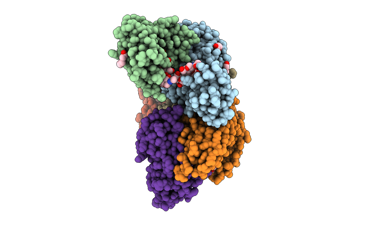
5MOI image
Entry Detail
PDB ID:
5MOI
Keywords:
Title:
Crystal structure of human IgE-Fc epsilon 3-4
Biological Source:
Source Organism:
Host Organism:
PDB Version:
Deposition Date:
2016-12-14
Release Date:
2018-01-10
Method Details:
Experimental Method:
Resolution:
2.20 Å
R-Value Free:
0.23
R-Value Work:
0.21
R-Value Observed:
0.21
Space Group:
P 1
Macromolecular Entities
Polymer Type:polypeptide(L)
Description:Ig epsilon chain C region
Chain IDs:A, B, C, D, E, F
Chain Length:223
Number of Molecules:6
Biological Source:Homo sapiens
Primary Citation
Thermal sensitivity and flexibility of the C epsilon 3 domains in immunoglobulin E.
Biochim. Biophys. Acta 1865 1336 1347 (2017)
PMID: 28844738 DOI: 10.1016/j.bbapap.2017.08.005

Abstact

Immunoglobulin E (IgE) is the antibody that plays a central role in the mechanisms of allergic diseases such as asthma. Interactions with its receptors, FcεRI on mast cells and CD23 on B cells, are mediated by the Fc region, a dimer of the Cε2, Cε3 and Cε4 domains. A sub-fragment lacking the Cε2 domains, Fcε3-4, also binds to both receptors, although receptor binding almost exclusively involves the Cε3 domains. This domain also contains the N-linked glycosylation site conserved in other isotypes. We report here the crystal structures of IgE-Fc and Fcε3-4 at the highest resolutions yet determined, 1.75Å and 2.0Å respectively, revealing unprecedented detail regarding the carbohydrate and its interactions with protein domains. Analysis of the crystallographic B-factors of these, together with all earlier IgE-Fc and Fcε3-4 structures, shows that the Cε3 domains exhibit the greatest intrinsic flexibility and quaternary structural variation within IgE-Fc. Intriguingly, both well-ordered carbohydrate and disordered polypeptide can be seen within the same Cε3 domain. A simplified method for comparing the quaternary structures of the Cε3 domains in free and receptor-bound IgE-Fc structures is presented, which clearly delineates the FcεRI and CD23 bound states. Importantly, differential scanning fluorimetric analysis of IgE-Fc and Fcε3-4 identifies Cε3 as the domain most susceptible to thermally-induced unfolding, and responsible for the characteristically low melting temperature of IgE.

Legend

Protein

Chemical

Disease

Primary Citation of related structures