Deposition Date
2016-12-13
Release Date
2017-01-11
Last Version Date
2024-05-08
Entry Detail
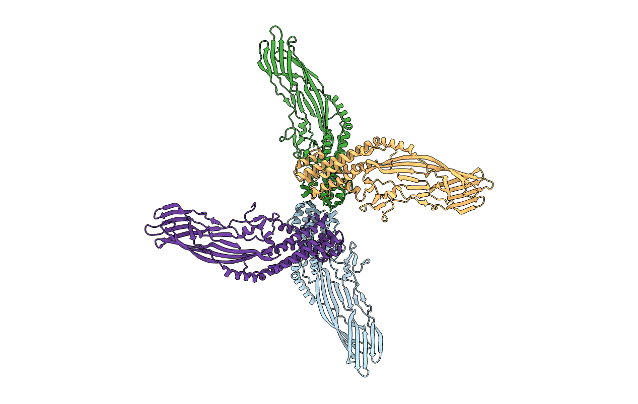
Biological Source:
Source Organism(s):
Enterobacteria phage Qbeta (Taxon ID: 39803)
Expression System(s):
Method Details:
Experimental Method:
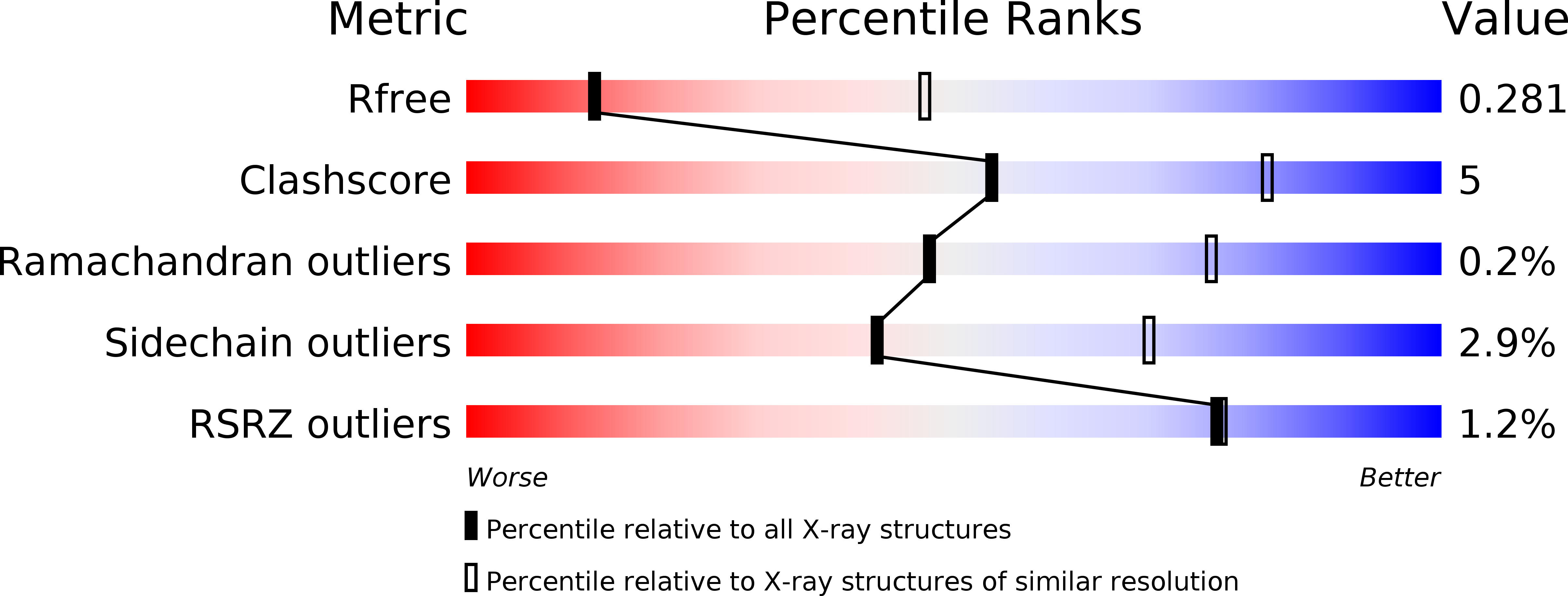
Resolution:
3.32 Å
R-Value Free:
0.27
R-Value Work:
0.22
R-Value Observed:
0.23
Space Group:
C 2 2 21