5MK6 image
Deposition Date 2016-12-02
Release Date 2018-03-28
Last Version Date 2024-05-08
Entry Detail
PDB ID:
5MK6
Keywords:
Title:
Crystal structure of the receptor-binding domain of botulinum neurotoxin A1 (crystal form 1)
Biological Source:
Source Organism:
Host Organism:
Method Details:
Experimental Method:
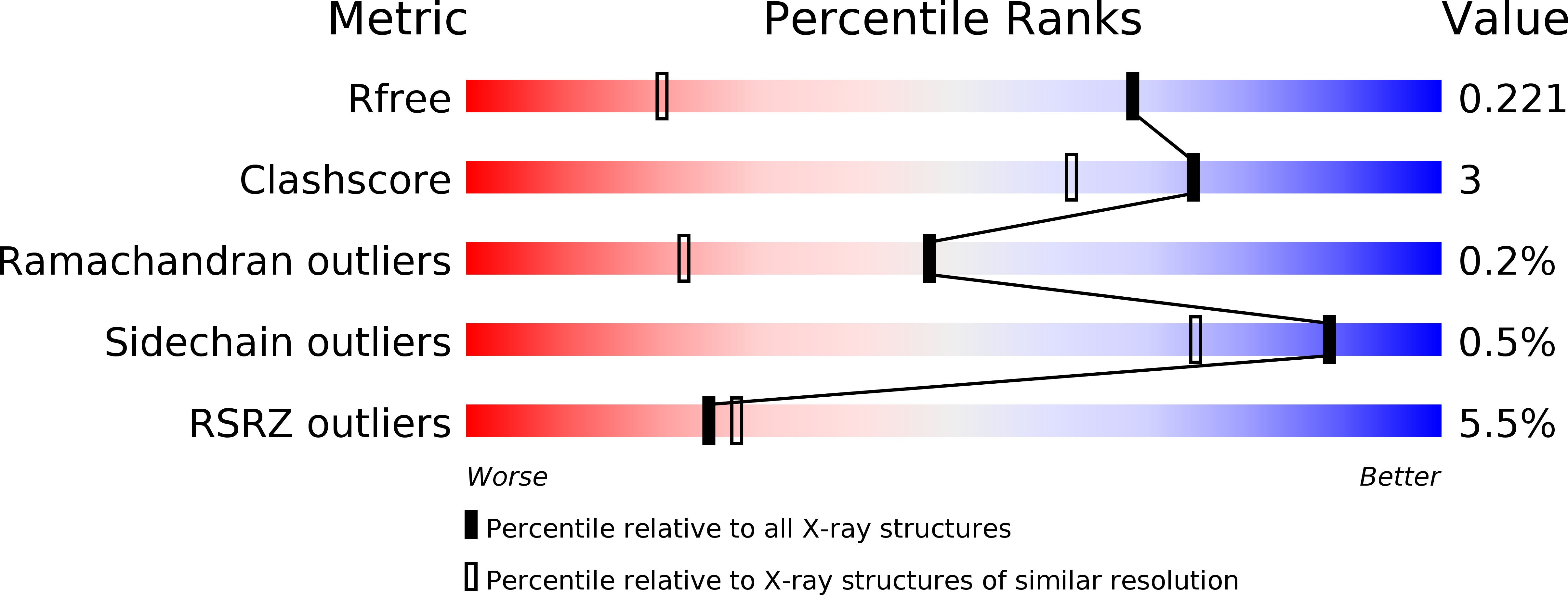
Resolution:
1.45 Å
R-Value Free:
0.22
R-Value Work:
0.17
R-Value Observed:
0.17
Space Group:
P 21 21 21
Macromolecular Entities
Structures with similar UniProt ID
Protein Blast
Polymer Type:polypeptide(L)
Molecule:Botulinum neurotoxin type A
Chain IDs:A
Chain Length:433
Number of Molecules:1
Biological Source:Clostridium botulinum
Ligand Molecules
Primary Citation
High resolution crystal structures of the receptor-binding domain ofClostridium botulinumneurotoxin serotypes A and FA.
PeerJ 6 e4552 e4552 (2018)
PMID: 29576992 DOI: 10.7717/peerj.4552

Abstact

The binding specificity of botulinum neurotoxins (BoNTs) is primarily a consequence of their ability to bind to multiple receptors at the same time. BoNTs consist of three distinct domains, a metalloprotease light chain (LC), a translocation domain (HN) and a receptor-binding domain (HC). Here we report the crystal structure of HC/FA, complementing an existing structure through the modelling of a previously unresolved loop which is important for receptor-binding. Our HC/FA structure also contains a previously unidentified disulphide bond, which we have also observed in one of two crystal forms of HC/A1. This may have implications for receptor-binding and future recombinant toxin production.

Legend

Protein

Chemical

Disease

Primary Citation of related structures