5MIX image
Deposition Date 2016-11-29
Release Date 2017-03-29
Last Version Date 2024-11-13
Entry Detail
PDB ID:
5MIX
Keywords:
Title:
Extracellular domain of human CD83 - trigonal crystal form
Biological Source:
Source Organism:
Homo sapiens (Taxon ID: 9606)
Method Details:
Experimental Method:
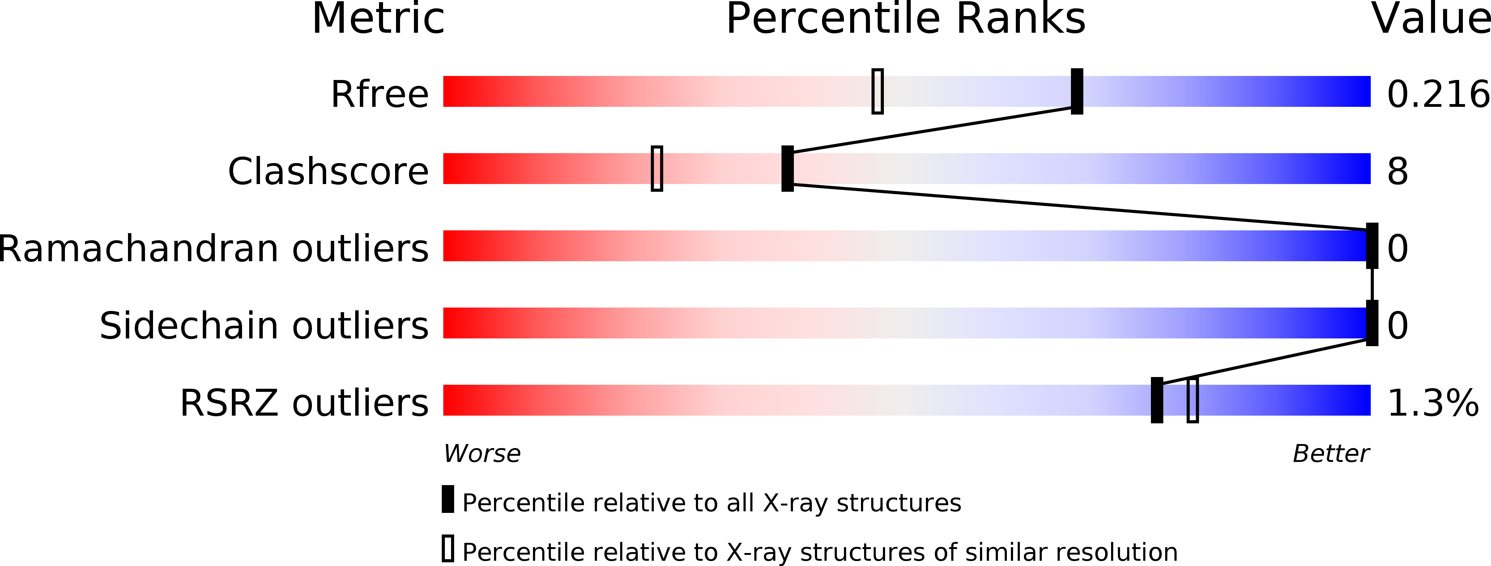
Resolution:
1.70 Å
R-Value Free:
0.21
R-Value Work:
0.18
R-Value Observed:
0.19
Space Group:
P 3 2 1
Macromolecular Entities
Polymer Type:polypeptide(L)
Molecule:CD83 antigen
Gene (Uniprot):CD83
Mutations:C27S, C100S, C129S
Chain IDs:A
Chain Length:116
Number of Molecules:1
Biological Source:Homo sapiens
Primary Citation
Crystal Structure of the Extracellular Domain of the Human Dendritic Cell Surface Marker CD83.
J. Mol. Biol. 429 1227 1243 (2017)
PMID: 28315353 DOI: 10.1016/j.jmb.2017.03.009

Abstact

CD83 is a type-I membrane protein and an efficient marker for identifying mature dendritic cells. Whereas membrane-bound, full-length CD83 co-stimulates the immune system, a soluble variant (sCD83), consisting of the extracellular domain only, displays strong immune-suppressive activities. Besides a prediction that sCD83 adopts a V-set Ig-like fold, however, little is known about the molecular architecture of CD83 and the mechanism by which CD83 exerts its function on dendritic cells and additional immune cells. Here, we report the crystal structure of human sCD83 up to a resolution of 1.7Å solved in three different crystal forms. Interestingly, β-strands C', C″, and D that are typical for V-set Ig-domains could not be traced in sCD83. Mass spectrometry analyses, limited proteolysis experiments, and bioinformatics studies show that the corresponding segment displays enhanced main-chain accessibility, extraordinary low sequence conservation, and a predicted high disorder propensity. Chimeric proteins with amino acid swaps in this segment show unaltered immune-suppressive activities in a TNF-α assay when compared to wild-type sCD83. This strongly indicates that this segment does not participate in the biological activity of CD83. The crystal structure of CD83 shows the recurrent formation of dimers and trimers in the various crystal forms and reveals strong structural similarities between sCD83 and B7 family members and CD48, a signaling lymphocyte activation molecule family member. This suggests that CD83 exerts its immunological activity by mixed homotypic and heterotypic interactions as typically observed for proteins present in the immunological synapse.

Legend

Protein

Chemical

Disease

Primary Citation of related structures