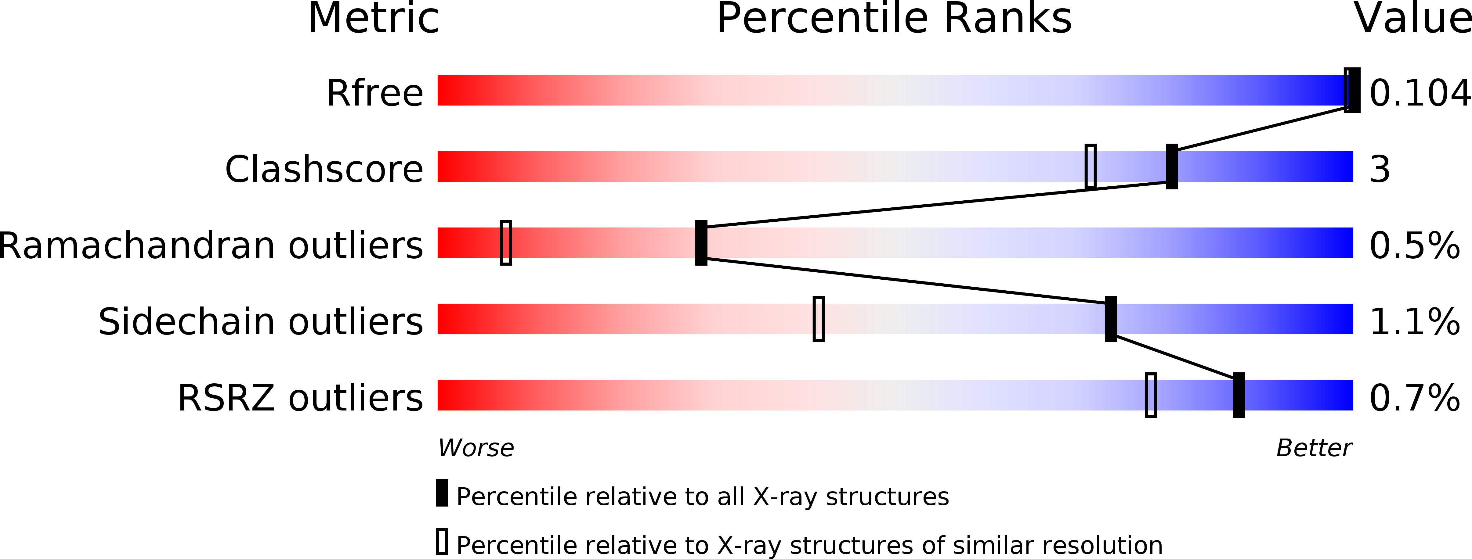
5MEH image
Deposition Date 2016-11-14
Release Date 2016-12-21
Last Version Date 2024-05-08
Entry Detail
PDB ID:
5MEH
Keywords:
Title:
Crystal structure of alpha-1,2-mannosidase from Caulobacter K31 strain in complex with 1-deoxymannojirimycin
Biological Source:
Source Organism:
Host Organism:
Method Details:
Experimental Method:
Resolution:
0.95 Å
R-Value Free:
0.10
R-Value Work:
0.09
R-Value Observed:
0.09
Space Group:
H 3
Macromolecular Entities
Structures with similar UniProt ID
Protein Blast
Polymer Type:polypeptide(L)
Molecule:Mannosyl-oligosaccharide 1,2-alpha-mannosidase
Gene (Uniprot):Caul_4035
Chain IDs:A
Chain Length:447
Number of Molecules:1
Biological Source:Caulobacter sp.
Primary Citation
Conformational Behaviour of Azasugars Based on Mannuronic Acid.
Chembiochem 18 1297 1304 (2017)
PMID: 28256791 DOI: 10.1002/cbic.201700080

Abstact

A set of mannuronic-acid-based iminosugars, consisting of the C-5-carboxylic acid, methyl ester and amide analogues of 1deoxymannorjirimicin (DMJ), was synthesised and their pH-dependent conformational behaviour was studied. Under acidic conditions the methyl ester and the carboxylic acid adopted an "inverted" 1 C4 chair conformation as opposed to the "normal" 4 C1 chair at basic pH. This conformational change is explained in terms of the stereoelectronic effects of the ring substituents and it parallels the behaviour of the mannuronic acid ester oxocarbenium ion. Because of this solution-phase behaviour, the mannuronic acid ester azasugar was examined as an inhibitor for a Caulobacter GH47 mannosidase that hydrolyses its substrates by way of a reaction itinerary that proceeds through a 3 H4 transition state. No binding was observed for the mannuronic acid ester azasugar, but sub-atomic resolution data were obtained for the DMJ⋅CkGH47 complex, showing two conformations-3 S1 and 1 C4 -for the DMJ inhibitor.

Legend

Protein

Chemical

Disease

Primary Citation of related structures