5MDJ image
Deposition Date 2016-11-11
Release Date 2018-02-21
Last Version Date 2024-11-20
Entry Detail
PDB ID:
5MDJ
Keywords:
Title:
Crystal structure of an O2-tolerant [NiFe]-hydrogenase from Ralstonia eutropha in a its as-isolated high-pressurized form
Biological Source:
Source Organism:
Host Organism:
Method Details:
Experimental Method:
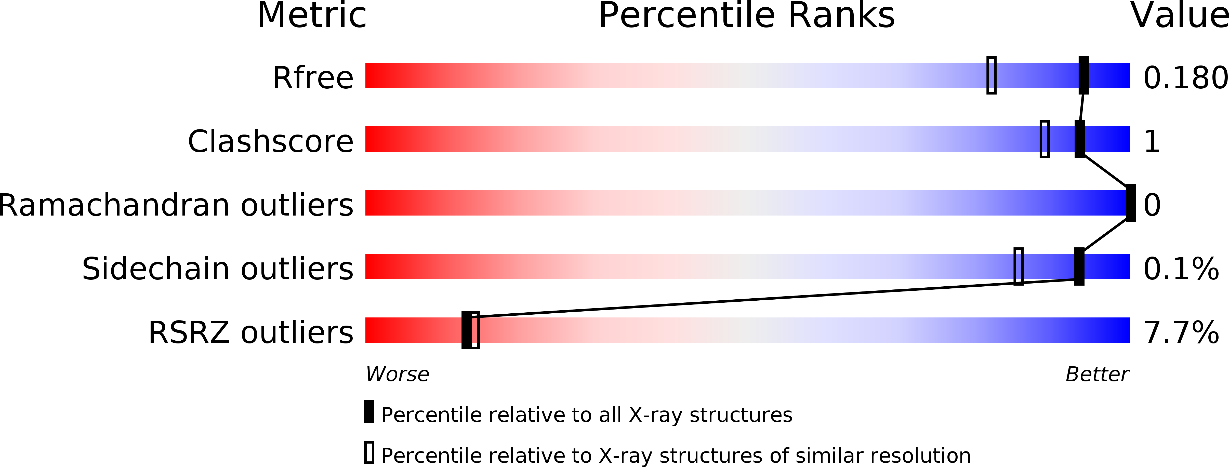
Resolution:
1.48 Å
R-Value Free:
0.17
R-Value Work:
0.14
R-Value Observed:
0.14
Space Group:
P 21 21 21
Macromolecular Entities
Structures with similar UniProt ID
Protein Blast
Polymer Type:polypeptide(L)
Molecule:Uptake hydrogenase large subunit; HOXG
Gene (Uniprot):hoxG
Chain IDs:A (auth: L)
Chain Length:603
Number of Molecules:1
Biological Source:Ralstonia eutropha H16
Structures with similar UniProt ID
Protein Blast
Polymer Type:polypeptide(L)
Molecule:Uptake hydrogenase small subunit; HOXK
Gene (Uniprot):hoxK
Chain IDs:B (auth: S)
Chain Length:328
Number of Molecules:1
Biological Source:Ralstonia eutropha H16
Primary Citation
Tracking the route of molecular oxygen in O2-tolerant membrane-bound [NiFe] hydrogenase.
Proc. Natl. Acad. Sci. U.S.A. 115 E2229 E2237 (2018)
PMID: 29463722 DOI: 10.1073/pnas.1712267115

Abstact

[NiFe] hydrogenases catalyze the reversible splitting of H(2) into protons and electrons at a deeply buried active site. The catalytic center can be accessed by gas molecules through a hydrophobic tunnel network. While most [NiFe] hydrogenases are inactivated by O(2), a small subgroup, including the membrane-bound [NiFe] hydrogenase (MBH) of Ralstonia eutropha, is able to overcome aerobic inactivation by catalytic reduction of O(2) to water. This O(2) tolerance relies on a special [4Fe3S] cluster that is capable of releasing two electrons upon O(2) attack. Here, the O(2) accessibility of the MBH gas tunnel network has been probed experimentally using a "soak-and-freeze" derivatization method, accompanied by protein X-ray crystallography and computational studies. This combined approach revealed several sites of O(2) molecules within a hydrophobic tunnel network leading, via two tunnel entrances, to the catalytic center of MBH. The corresponding site occupancies were related to the O(2) concentrations used for MBH crystal derivatization. The examination of the O(2)-derivatized data furthermore uncovered two unexpected structural alterations at the [4Fe3S] cluster, which might be related to the O(2) tolerance of the enzyme.

Legend

Protein

Chemical

Disease

Primary Citation of related structures