Deposition Date
2016-11-09
Release Date
2017-12-20
Last Version Date
2024-11-06
Entry Detail
PDB ID:
5MC0
Keywords:
Title:
Crystal Structure of delTyr231 mutant of Human Prolidase with Mn ions and GlyPro ligand
Biological Source:
Source Organism(s):
Homo sapiens (Taxon ID: 9606)
Expression System(s):
Method Details:
Experimental Method:
Resolution:
1.56 Å
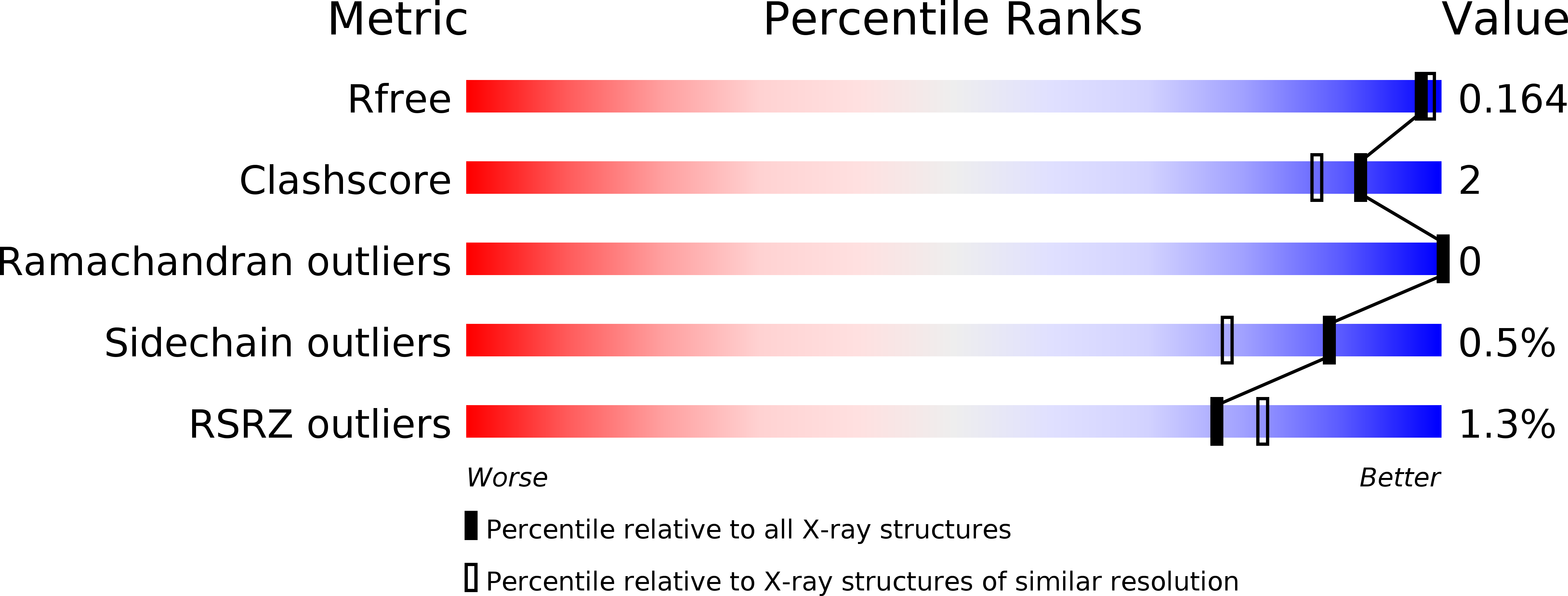
R-Value Free:
0.15
R-Value Work:
0.11
R-Value Observed:
0.11
Space Group:
C 2 2 21