5MAP image
Deposition Date 2016-11-04
Release Date 2017-05-10
Last Version Date 2024-01-17
Entry Detail
PDB ID:
5MAP
Keywords:
Title:
X-ray generated oxyferrous complex of DtpA from Streptomyces lividans
Biological Source:
Source Organism:
Method Details:
Experimental Method:
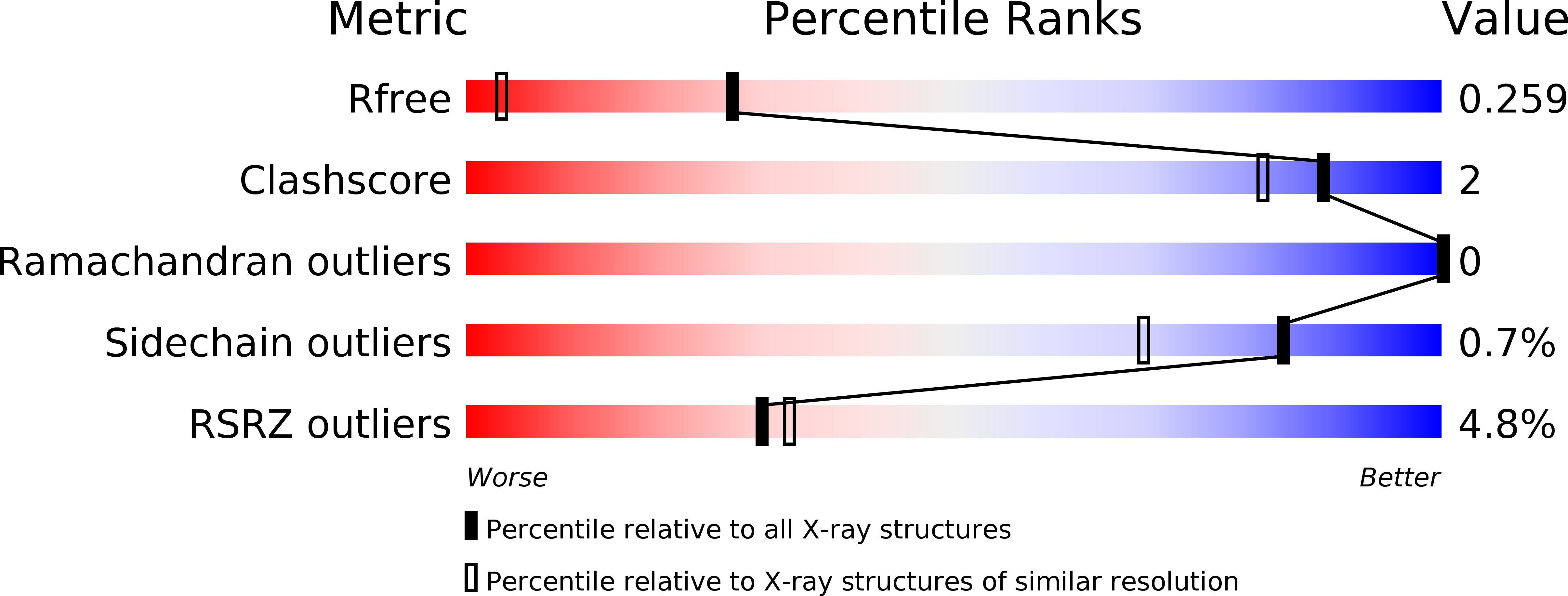
Resolution:
1.49 Å
R-Value Free:
0.25
R-Value Work:
0.21
R-Value Observed:
0.22
Space Group:
P 1 21 1
Macromolecular Entities
Polymer Type:polypeptide(L)
Molecule:DtpA
Chain IDs:A, B
Chain Length:376
Number of Molecules:2
Biological Source:Streptomyces lividans TK24
Primary Citation
Photoreduction and validation of haem-ligand intermediate states in protein crystals by in situ single-crystal spectroscopy and diffraction.
IUCrJ 4 263 270 (2017)
PMID: 28512573 DOI: 10.1107/S2052252517002159

Abstact

Powerful synergies are available from the combination of multiple methods to study proteins in the crystalline form. Spectroscopies which probe the same region of the crystal from which X-ray crystal structures are determined can give insights into redox, ligand and spin states to complement the information gained from the electron-density maps. The correct assignment of crystal structures to the correct protein redox and ligand states is essential to avoid the misinterpretation of structural data. This is a particular concern for haem proteins, which can occupy a wide range of redox states and are exquisitely sensitive to becoming reduced by solvated electrons generated from interactions of X-rays with water molecules in the crystal. Here, single-crystal spectroscopic fingerprinting has been applied to investigate the laser photoreduction of ferric haem in cytochrome c'. Furthermore, in situ X-ray-driven generation of haem intermediates in crystals of the dye-decolourizing-type peroxidase A (DtpA) from Streptomyces lividans is described.

Legend

Protein

Chemical

Disease

Primary Citation of related structures