5MAC image
Entry Detail
PDB ID:
5MAC
Keywords:
Title:
Crystal structure of decameric Methanococcoides burtonii Rubisco complexed with 2-carboxyarabinitol bisphosphate
Biological Source:
Source Organism:
Host Organism:
PDB Version:
Deposition Date:
2016-11-03
Release Date:
2017-02-08
Method Details:
Experimental Method:
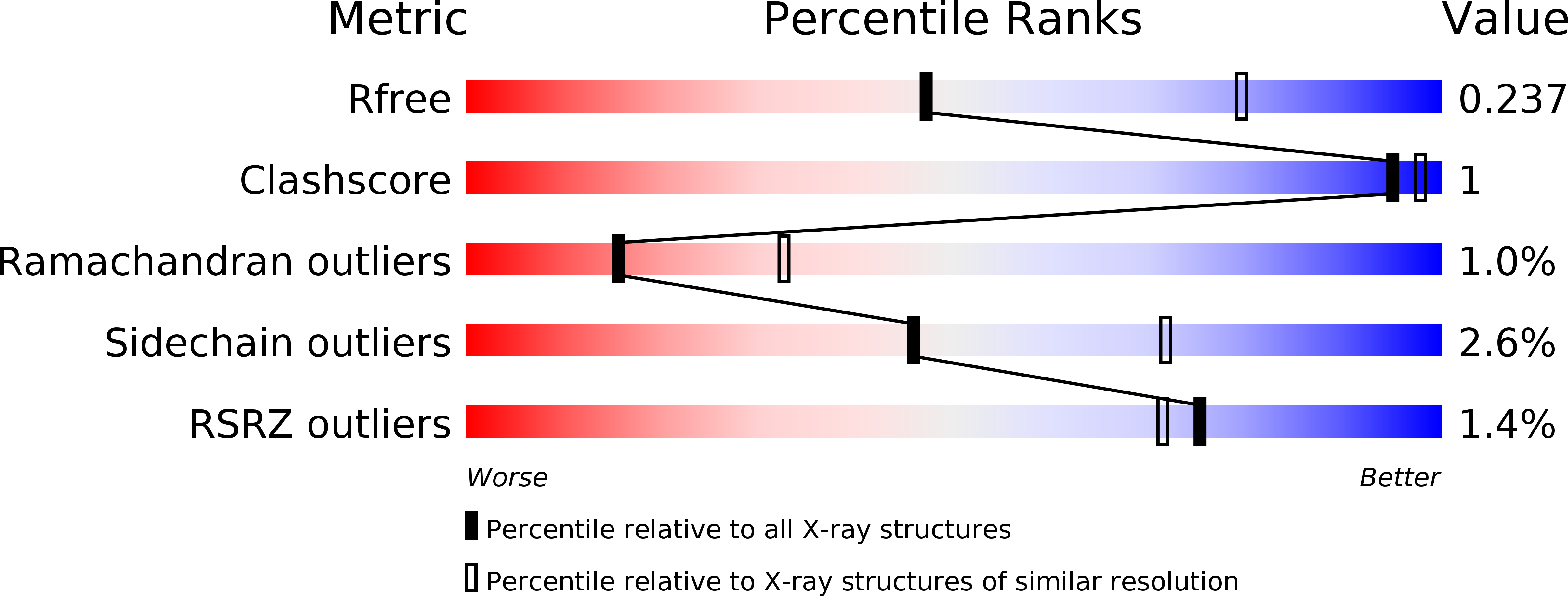
Resolution:
2.60 Å
R-Value Free:
0.22
R-Value Work:
0.18
R-Value Observed:
0.19
Space Group:
P 3 2 1
Macromolecular Entities
Polymer Type:polypeptide(L)
Description:Ribulose-1,5-bisphosphate carboxylase-oxygenase type III
Chain IDs:A, B, C, D, E
Chain Length:474
Number of Molecules:5
Biological Source:Methanococcoides burtonii
Modified Residue
Compound ID Chain ID Parent Comp ID Details 2D Image
KCX A LYS modified residue
Primary Citation
A unique structural domain in Methanococcoides burtonii ribulose-1,5-bisphosphate carboxylase/oxygenase (Rubisco) acts as a small subunit mimic.
J. Biol. Chem. 292 6838 6850 (2017)
PMID: 28154188 DOI: 10.1074/jbc.M116.767145

Abstact

The catalytic inefficiencies of the CO2-fixing enzyme ribulose-1,5-bisphosphate carboxylase/oxygenase (Rubisco) often limit plant productivity. Strategies to engineer more efficient plant Rubiscos have been hampered by evolutionary constraints, prompting interest in Rubisco isoforms from non-photosynthetic organisms. The methanogenic archaeon Methanococcoides burtonii contains a Rubisco isoform that functions to scavenge the ribulose-1,5-bisphosphate (RuBP) by-product of purine/pyrimidine metabolism. The crystal structure of M. burtonii Rubisco (MbR) presented here at 2.6 Å resolution is composed of catalytic large subunits (LSu) assembled into pentamers of dimers, (L2)5, and differs from Rubiscos from higher plants where LSus are glued together by small subunits (SSu) into hexadecameric L8S8 enzymes. MbR contains a unique 29-amino acid insertion near the C terminus, which folds as a separate domain in the structure. This domain, which is visualized for the first time in this study, is located in a similar position to SSus in L8S8 enzymes between LSus of adjacent L2 dimers, where negatively charged residues coordinate around a Mg2+ ion in a fashion that suggests this domain may be important for the assembly process. The Rubisco assembly domain is thus an inbuilt SSu mimic that concentrates L2 dimers. MbR assembly is ligand-stimulated, and we show that only 6-carbon molecules with a particular stereochemistry at the C3 carbon can induce oligomerization. Based on MbR structure, subunit arrangement, sequence, phylogenetic distribution, and function, MbR and a subset of Rubiscos from the Methanosarcinales order are proposed to belong to a new Rubisco subgroup, named form IIIB.

Legend

Protein

Chemical

Disease

Primary Citation of related structures