5M6D image
Deposition Date 2016-10-25
Release Date 2017-01-11
Last Version Date 2024-01-17
Entry Detail
PDB ID:
5M6D
Keywords:
Title:
Streptococcus pneumoniae Glyceraldehyde-3-Phosphate Dehydrogenase (SpGAPDH) crystal structure
Biological Source:
Source Organism:
Host Organism:
Method Details:
Experimental Method:
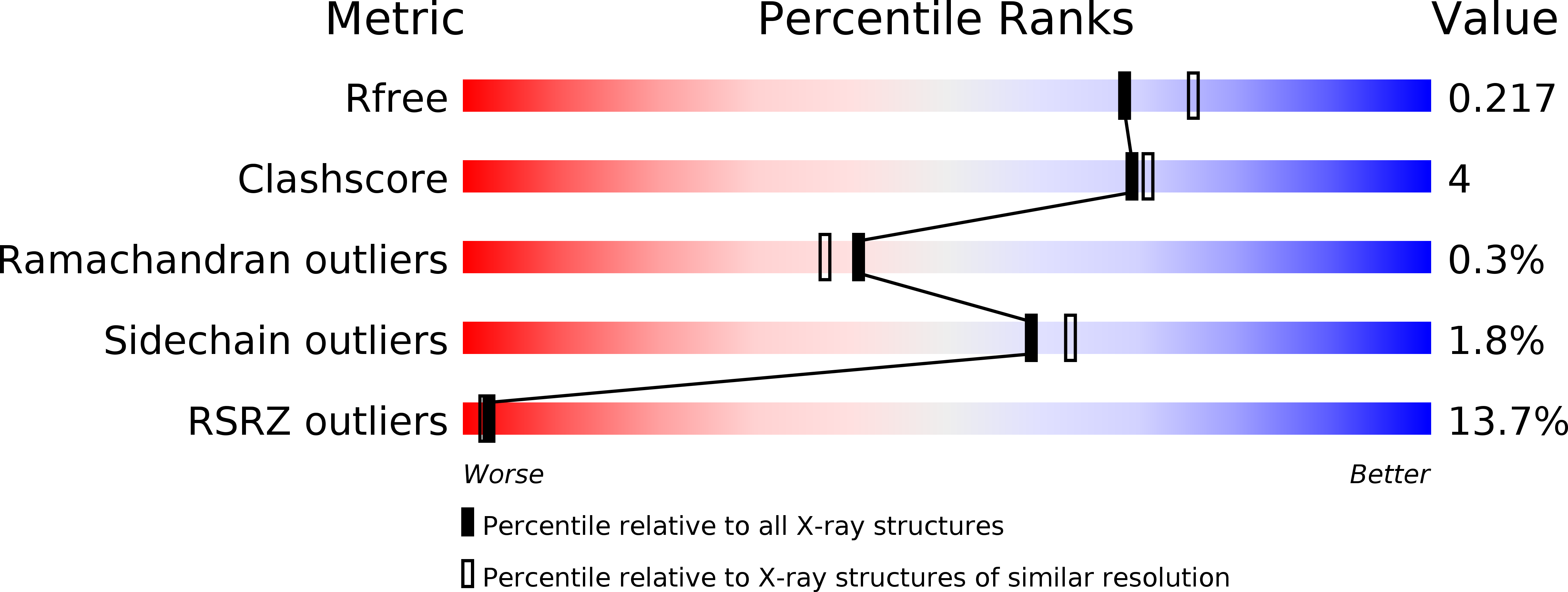
Resolution:
2.00 Å
R-Value Free:
0.21
R-Value Work:
0.19
R-Value Observed:
0.19
Space Group:
C 1 2 1
Macromolecular Entities
Structures with similar UniProt ID
Protein Blast
Polymer Type:polypeptide(L)
Molecule:Glyceraldehyde-3-phosphate dehydrogenase
Gene (Uniprot):gapdh
Chain IDs:A, B
Chain Length:355
Number of Molecules:2
Biological Source:Streptococcus pneumoniae
Modified Residue
Compound ID Chain ID Parent Comp ID Details 2D Image
CSU A CYS modified residue
Primary Citation
Deciphering Key Residues Involved in the Virulence-promoting Interactions between Streptococcus pneumoniae and Human Plasminogen.
J. Biol. Chem. 292 2217 2225 (2017)
PMID: 28011643 DOI: 10.1074/jbc.M116.764209

Abstact

Bacterial pathogens recruit circulating proteins to their own surfaces, co-opting the host protein functions as a mechanism of virulence. Particular attention has focused on the binding of plasminogen (Plg) to bacterial surfaces, as it has been shown that this interaction contributes to bacterial adhesion to host cells, invasion of host tissues, and evasion of the immune system. Several bacterial proteins are known to serve as receptors for Plg including glyceraldehyde-3-phosphate dehydrogenase (GAPDH), a cytoplasmic enzyme that appears on the cell surface in this moonlighting role. Although Plg typically binds to these receptors via several lysine-binding domains, the specific interactions that occur have not been documented in all cases. However, identification of the relevant residues could help define strategies for mitigating the virulence of important human pathogens, such as Streptococcus pneumoniae (Sp). To shed light on this question, we have described a combination of peptide-spot array screening, competition and SPR assays, high-resolution crystallography, and mutational analyses to characterize the interaction between SpGAPDH and Plg. We identified three SpGAPDH lysine residues that were instrumental in defining the kinetic and thermodynamic parameters of the interaction. Altogether, the integration of the data presented in this work allows us to propose a structural model for the molecular interaction of the SpGAPDH-Plg complex.

Legend

Protein

Chemical

Disease

Primary Citation of related structures