5M63 image
Deposition Date 2016-10-24
Release Date 2017-05-03
Last Version Date 2024-11-06
Entry Detail
PDB ID:
5M63
Keywords:
Title:
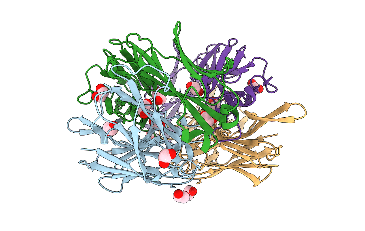
Crystal structure of group B Streptococcus type III DP2 oligosaccharide bound to Fab NVS-1-19-5
Biological Source:
Source Organism:
Host Organism:
Method Details:
Experimental Method:
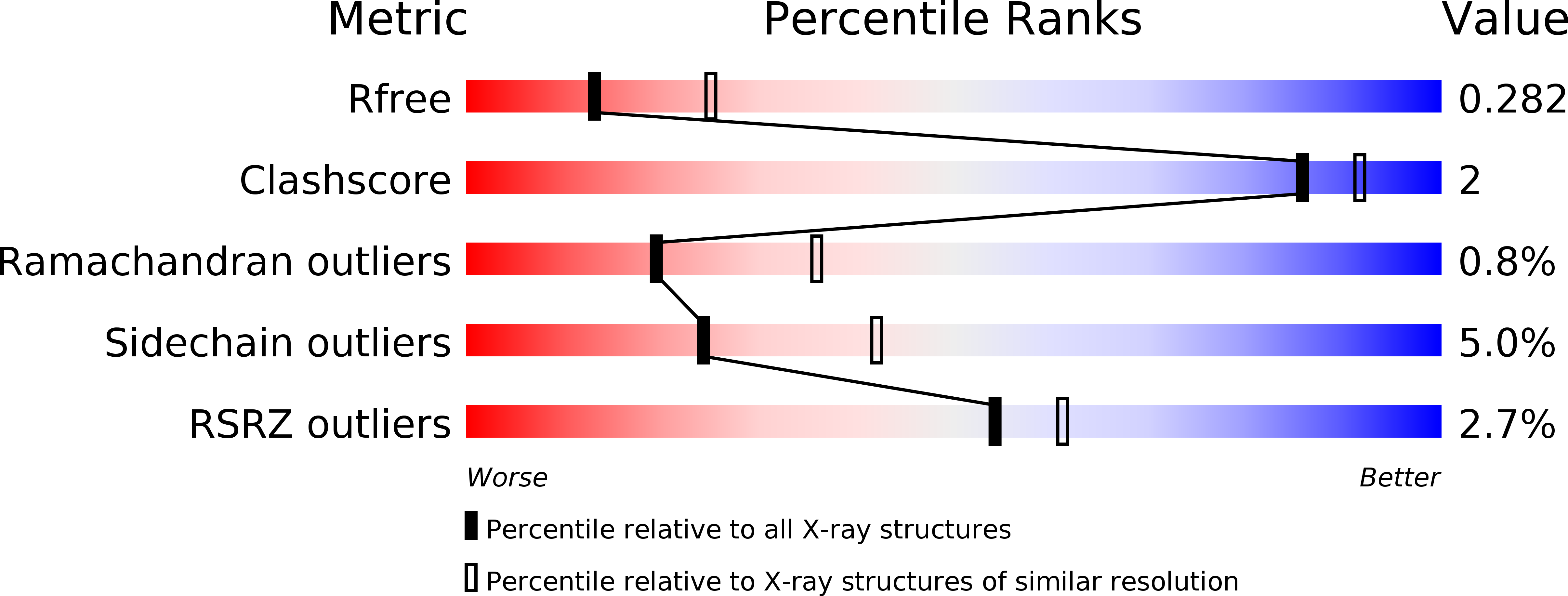
Resolution:
2.74 Å
R-Value Free:
0.28
R-Value Work:
0.23
R-Value Observed:
0.23
Space Group:
C 2 2 21
Macromolecular Entities
Polymer Type:polypeptide(L)
Molecule:H chain of Fab NVS-1-19-5
Chain IDs:A (auth: H), C (auth: M)
Chain Length:248
Number of Molecules:2
Biological Source:Oryctolagus cuniculus
Polymer Type:polypeptide(L)
Molecule:L chain of Fab NVS-1-19-5
Chain IDs:B (auth: L), D (auth: N)
Chain Length:239
Number of Molecules:2
Biological Source:Oryctolagus cuniculus
Primary Citation
Structure of a protective epitope of group B Streptococcus type III capsular polysaccharide.
Proc. Natl. Acad. Sci. U.S.A. 114 5017 5022 (2017)
PMID: 28439022 DOI: 10.1073/pnas.1701885114

Abstact

Despite substantial progress in the prevention of group B Streptococcus (GBS) disease with the introduction of intrapartum antibiotic prophylaxis, this pathogen remains a leading cause of neonatal infection. Capsular polysaccharide conjugate vaccines have been tested in phase I/II clinical studies, showing promise for further development. Mapping of epitopes recognized by protective antibodies is crucial for understanding the mechanism of action of vaccines and for enabling antigen design. In this study, we report the structure of the epitope recognized by a monoclonal antibody with opsonophagocytic activity and representative of the protective response against type III GBS polysaccharide. The structure and the atomic-level interactions were determined by saturation transfer difference (STD)-NMR and X-ray crystallography using oligosaccharides obtained by synthetic and depolymerization procedures. The GBS PSIII epitope is made by six sugars. Four of them derive from two adjacent repeating units of the PSIII backbone and two of them from the branched galactose-sialic acid disaccharide contained in this sequence. The sialic acid residue establishes direct binding interactions with the functional antibody. The crystal structure provides insight into the molecular basis of antibody-carbohydrate interactions and confirms that the conformational epitope is not required for antigen recognition. Understanding the structural basis of immune recognition of capsular polysaccharide epitopes can aid in the design of novel glycoconjugate vaccines.

Legend

Protein

Chemical

Disease

Primary Citation of related structures
Feedback Form
Name
Email
Institute
Feedback