5M4T image
Entry Detail
PDB ID:
5M4T
Title:
C-terminal domain structure of VSG M1.1
Biological Source:
Source Organism:
Host Organism:
PDB Version:
Deposition Date:
2016-10-19
Release Date:
2017-09-20
Method Details:
Experimental Method:
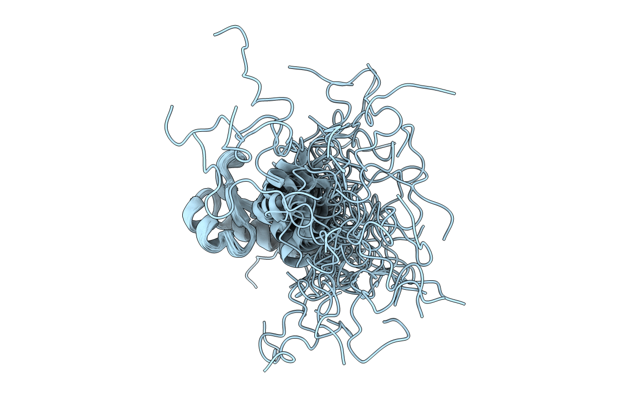
Conformers Calculated:
100
Conformers Submitted:
35
Selection Criteria:
structures with the lowest energy
Macromolecular Entities
Polymer Type:polypeptide(L)
Description:Variant surface glycoprotein MITAT 1.1
Chain IDs:A
Chain Length:73
Number of Molecules:1
Biological Source:Trypanosoma brucei brucei
Ligand Molecules
Primary Citation
Structural basis for the shielding function of the dynamic trypanosome variant surface glycoprotein coat.
Nat Microbiol 2 1523 1532 (2017)
PMID: 28894098 DOI: 10.1038/s41564-017-0013-6

Abstact

The most prominent defence of the unicellular parasite Trypanosoma brucei against the host immune system is a dense coat that comprises a variant surface glycoprotein (VSG). Despite the importance of the VSG family, no complete structure of a VSG has been reported. Making use of high-resolution structures of individual VSG domains, we employed small-angle X-ray scattering to elucidate the first two complete VSG structures. The resulting models imply that the linker regions confer great flexibility between domains, which suggests that VSGs can adopt two main conformations to respond to obstacles and changes of protein density, while maintaining a protective barrier at all times. Single-molecule diffusion measurements of VSG in supported lipid bilayers substantiate this possibility, as two freely diffusing populations could be detected. This translates into a highly flexible overall topology of the surface VSG coat, which displays both lateral movement in the plane of the membrane and variation in the overall thickness of the coat.

Legend

Protein

Chemical

Disease

Primary Citation of related structures