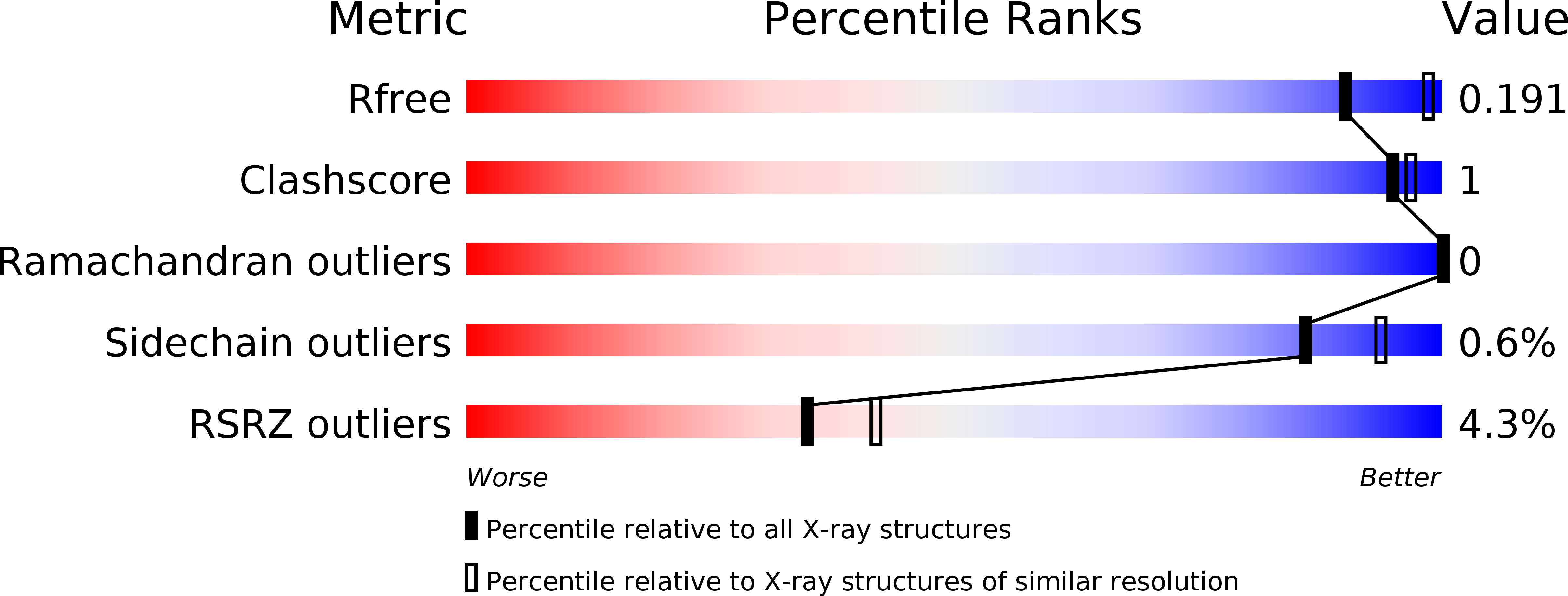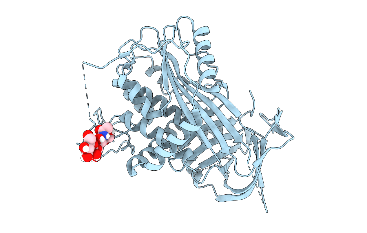
5M3Y image
Entry Detail
PDB ID:
5M3Y
Title:
Crystal structure of human glycosylated angiotensinogen
Biological Source:
Source Organism:
Host Organism:
PDB Version:
Deposition Date:
2016-10-17
Release Date:
2017-12-20
Method Details:
Experimental Method:
Resolution:
2.30 Å
R-Value Free:
0.18
R-Value Work:
0.16
R-Value Observed:
0.17
Space Group:
P 41
Macromolecular Entities
Polymer Type:polypeptide(L)
Description:Angiotensinogen
Mutations:N137Q, N271Q, N295Q, C232S, C308S
Chain IDs:A
Chain Length:458
Number of Molecules:1
Biological Source:Homo sapiens
Primary Citation
Structural basis for the specificity of renin-mediated angiotensinogen cleavage.
J. Biol. Chem. 294 2353 2364 (2019)
PMID: 30563843 DOI: 10.1074/jbc.RA118.006608

Abstact

The renin-angiotensin cascade is a hormone system that regulates blood pressure and fluid balance. Renin-mediated cleavage of the angiotensin I peptide from the N terminus of angiotensinogen (AGT) is the rate-limiting step of this cascade; however, the detailed molecular mechanism underlying this step is unclear. Here, we solved the crystal structures of glycosylated human AGT (2.30 Å resolution), its encounter complex with renin (2.55 Å), AGT cleaved in its reactive center loop (RCL; 2.97 Å), and spent AGT from which the N-terminal angiotensin peptide was removed (2.63 Å). These structures revealed that AGT undergoes profound conformational changes and binds renin through a tail-into-mouth allosteric mechanism that inserts the N terminus into a pocket equivalent to a hormone-binding site on other serpins. These changes fully extended the N-terminal tail, with the scissile bond for angiotensin release docked in renin's active site. Insertion of the N terminus into this pocket accompanied a complete unwinding of helix H of AGT, which, in turn, formed key interactions with renin in the complementary binding interface. Mutagenesis and kinetic analyses confirmed that renin-mediated production of angiotensin I is controlled by interactions of amino acid residues and glycan components outside renin's active-site cleft. Our findings indicate that AGT adapts unique serpin features for hormone delivery and binds renin through concerted movements in the N-terminal tail and in its main body to modulate angiotensin release. These insights provide a structural basis for the development of agents that attenuate angiotensin release by targeting AGT's hormone binding pocket.

Legend

Protein

Chemical

Disease

Primary Citation of related structures