5M26 image
Entry Detail
PDB ID:
5M26
Keywords:
Title:
Crystal structure of hydroquinone 1,2-dioxygenase from Sphingomonas sp. TTNP3 in complex with methylhydroquinone
Biological Source:
Source Organism:
PDB Version:
Deposition Date:
2016-10-12
Release Date:
2017-09-27
Method Details:
Experimental Method:
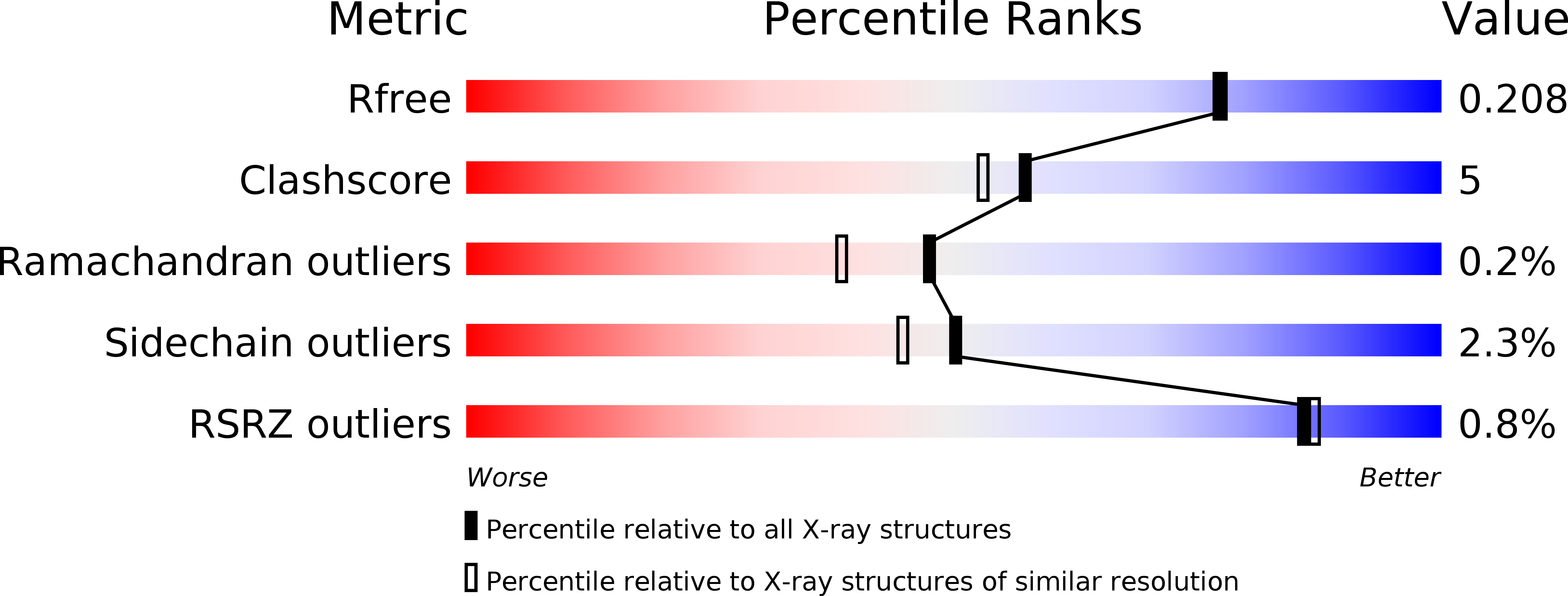
Resolution:
1.90 Å
R-Value Free:
0.20
R-Value Work:
0.16
R-Value Observed:
0.16
Space Group:
P 1 21 1
Macromolecular Entities
Polymer Type:polypeptide(L)
Description:Hydroquinone dioxygenase small subunit
Chain IDs:A, C, E, G
Chain Length:170
Number of Molecules:4
Biological Source:Sphingomonas sp. TTNP3
Polymer Type:polypeptide(L)
Description:Hydroquinone dioxygenase large subunit
Chain IDs:B, D, F, H
Chain Length:341
Number of Molecules:4
Biological Source:Sphingomonas sp. TTNP3
Primary Citation
The crystal structures of native hydroquinone 1,2-dioxygenase from Sphingomonas sp. TTNP3 and of substrate and inhibitor complexes.
Biochim. Biophys. Acta 1865 520 530 (2017)
PMID: 28232026 DOI: 10.1016/j.bbapap.2017.02.013

Abstact

The crystal structure of hydroquinone 1,2-dioxygenase, a Fe(II) ring cleaving dioxygenase from Sphingomonas sp. strain TTNP3, which oxidizes a wide range of hydroquinones to the corresponding 4-hydroxymuconic semialdehydes, has been solved by Molecular Replacement, using the coordinates of PnpCD from Pseudomonas sp. strain WBC-3. The enzyme is a heterotetramer, constituted of two subunits α and two β of 19 and 38kDa, respectively. Both the two subunits fold as a cupin, but that of the small α subunit lacks a competent metal binding pocket. Two tetramers are present in the asymmetric unit. Each of the four β subunits in the asymmetric unit binds one Fe(II) ion. The iron ion in each β subunit is coordinated to three protein residues, His258, Glu264, and His305 and a water molecule. The crystal structures of the complexes with the substrate methylhydroquinone, obtained under anaerobic conditions, and with the inhibitors 4-hydroxybenzoate and 4-nitrophenol were also solved. The structures of the native enzyme and of the complexes present significant differences in the active site region compared to PnpCD, the other hydroquinone 1,2-dioxygenase of known structure, and in particular they show a different coordination at the metal center.

Legend

Protein

Chemical

Disease

Primary Citation of related structures