5M1M image
Entry Detail
PDB ID:
5M1M
Keywords:
Title:
Crystal structure of matrix protein 1 from Influenza C virus (strain C/Ann Arbor/1/1950)
Biological Source:
PDB Version:
Deposition Date:
2016-10-09
Release Date:
2017-02-08
Method Details:
Experimental Method:
Resolution:
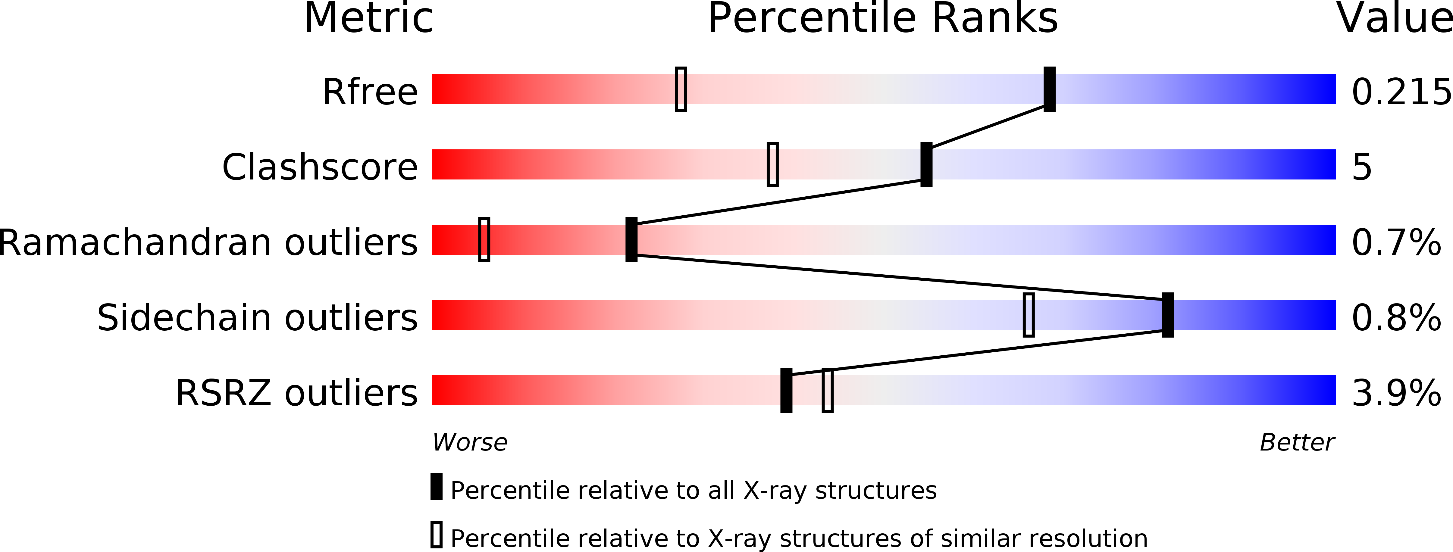
1.50 Å
R-Value Free:
0.21
R-Value Work:
0.18
R-Value Observed:
0.18
Space Group:
C 1 2 1
Macromolecular Entities
Polymer Type:polypeptide(L)
Description:Matrix protein 1
Chain IDs:A
Chain Length:155
Number of Molecules:1
Biological Source:Influenza C virus (strain C/Ann Arbor/1/1950)
Ligand Molecules
Primary Citation
The Matrix protein M1 from influenza C virus induces tubular membrane invaginations in an in vitro cell membrane model.
Sci Rep 7 40801 40801 (2017)
PMID: 28120862 DOI: 10.1038/srep40801

Abstact

Matrix proteins from enveloped viruses play an important role in budding and stabilizing virus particles. In order to assess the role of the matrix protein M1 from influenza C virus (M1-C) in plasma membrane deformation, we have combined structural and in vitro reconstitution experiments with model membranes. We present the crystal structure of the N-terminal domain of M1-C and show by Small Angle X-Ray Scattering analysis that full-length M1-C folds into an elongated structure that associates laterally into ring-like or filamentous polymers. Using negatively charged giant unilamellar vesicles (GUVs), we demonstrate that M1-C full-length binds to and induces inward budding of membrane tubules with diameters that resemble the diameter of viruses. Membrane tubule formation requires the C-terminal domain of M1-C, corroborating its essential role for M1-C polymerization. Our results indicate that M1-C assembly on membranes constitutes the driving force for budding and suggest that M1-C plays a key role in facilitating viral egress.

Legend

Protein

Chemical

Disease

Primary Citation of related structures