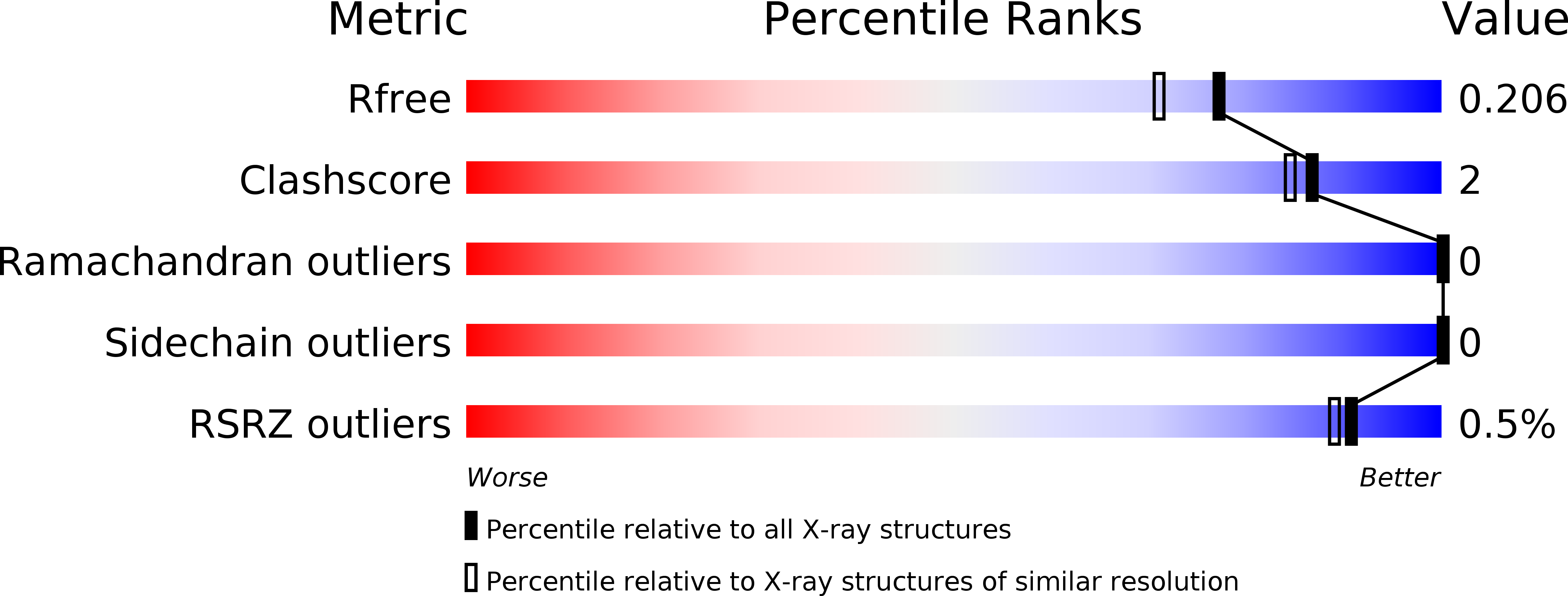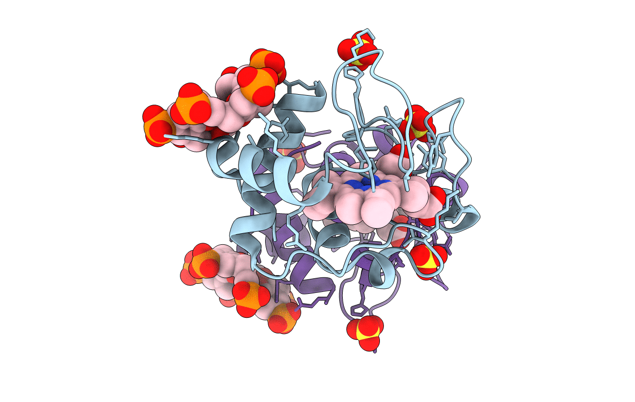
5LYC image
Deposition Date 2016-09-27
Release Date 2017-05-10
Last Version Date 2024-11-13
Entry Detail
PDB ID:
5LYC
Keywords:
Title:
Cytochrome c in complex with phosphonato-calix[6]arene
Biological Source:
Method Details:
Experimental Method:
Resolution:
1.80 Å
R-Value Free:
0.19
R-Value Work:
0.16
R-Value Observed:
0.17
Space Group:
P 43 21 2
Macromolecular Entities
Structures with similar UniProt ID
Protein Blast
Polymer Type:polypeptide(L)
Molecule:Cytochrome c iso-1
Gene (Uniprot):CYC1
Mutagens:C102T
Chain IDs:A, B
Chain Length:108
Number of Molecules:2
Biological Source:Saccharomyces cerevisiae (strain ATCC 204508 / S288c)
Primary Citation
Protein Dimerization on a Phosphonated Calix[6]arene Disc.
Angew. Chem. Int. Ed. Engl. 56 5517 5521 (2017)
PMID: 28407337 DOI: 10.1002/anie.201701500

Abstact

Complex formation between cationic cytochrome c and the water-soluble, poly-anionic p-phosphonatocalix[6]arene (pclx6) was investigated. A crystal structure (at 1.8 Å resolution) revealed a remarkable dimeric disc of pclx6 that acts like glue to mediate a symmetric (C2) protein dimer. The calixarene disc has a diameter of about 1.5 nm and masks about 360 Å2 of protein surface. The key protein-calixarene contacts occur via two linchpin lysines, with additional contacts provided by a small hydrophobic patch. The protein-calixarene supramolecular assemblies were observed in solution by size-exclusion chromatography with multi-angle light scattering and NMR spectroscopy. Using isothermal titration calorimetry and NMR data, an apparent Kd in the low micromolar range was determined for the charge-rich protein-calixarene complex. In contrast to p-sulfonatocalix[4]arene, the larger pclx6 has a single, well-defined binding site that mediates the assembly of cytochrome c in solution.

Legend

Protein

Chemical

Disease

Primary Citation of related structures