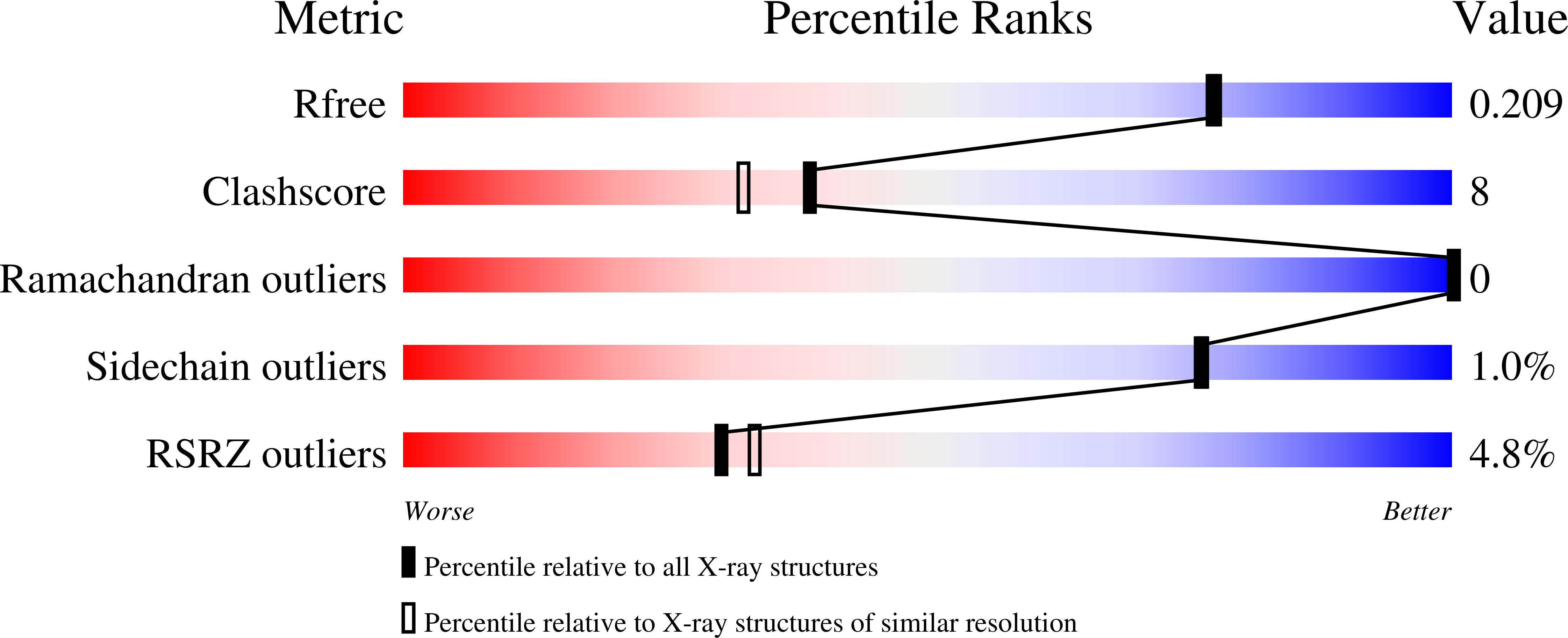
5LXT image
Deposition Date 2016-09-22
Release Date 2017-03-01
Last Version Date 2024-01-17
Entry Detail
PDB ID:
5LXT
Keywords:
Title:
Tubulin-Discodermolide complex
Biological Source:
Source Organism:
Rattus norvegicus (Taxon ID: 10116)
Gallus gallus (Taxon ID: 9031)
Bos taurus (Taxon ID: 9913)
Host Organism:
Method Details:
Experimental Method:
Resolution:
1.90 Å
R-Value Free:
0.20
R-Value Work:
0.17
R-Value Observed:
0.17
Space Group:
P 21 21 21
Macromolecular Entities
Structures with similar UniProt ID
Protein Blast
Polymer Type:polypeptide(L)
Molecule:Tubulin alpha-1B chain
Chain IDs:A, C
Chain Length:451
Number of Molecules:2
Biological Source:Bos taurus
Structures with similar UniProt ID
Protein Blast
Polymer Type:polypeptide(L)
Molecule:Tubulin beta-2B chain
Gene (Uniprot):TUBB2B
Chain IDs:B, D
Chain Length:445
Number of Molecules:2
Biological Source:Bos taurus
Structures with similar UniProt ID
Protein Blast
Polymer Type:polypeptide(L)
Molecule:Stathmin-4
Gene (Uniprot):Stmn4
Chain IDs:E
Chain Length:143
Number of Molecules:1
Biological Source:Rattus norvegicus
Polymer Type:polypeptide(L)
Molecule:TUBULIN-TYROSINE LIGASE
Chain IDs:F
Chain Length:384
Number of Molecules:1
Biological Source:Gallus gallus
Primary Citation
Structural Basis of Microtubule Stabilization by Discodermolide.
Chembiochem 18 905 909 (2017)
PMID: 28207984 DOI: 10.1002/cbic.201600696

Abstact

Microtubule-stabilizing agents (MSAs) are widely used in chemotherapy. Using X-ray crystallography we elucidated the detailed binding modes of two potent MSAs, (+)-discodermolide (DDM) and the DDM-paclitaxel hybrid KS-1-199-32, in the taxane pocket of β-tubulin. The two compounds bind in a very similar hairpin conformation, as previously observed in solution. However, they stabilize the M-loop of β-tubulin differently: KS-1-199-32 induces an M-loop helical conformation that is not observed for DDM. In the context of the microtubule structure, both MSAs connect the β-tubulin helices H6 and H7 and loop S9-S10 with the M-loop. This is similar to the structural effects elicited by epothilone A, but distinct from paclitaxel. Together, our data reveal differential binding mechanisms of DDM and KS-1-199-32 on tubulin.

Legend

Protein

Chemical

Disease

Primary Citation of related structures