5LXD image
Deposition Date 2016-09-20
Release Date 2016-10-26
Last Version Date 2024-11-06
Entry Detail
PDB ID:
5LXD
Keywords:
Title:
Crystal structure of DYRK2 in complex with EHT 1610 (compound 2)
Biological Source:
Source Organism:
Homo sapiens (Taxon ID: 9606)
Host Organism:
Method Details:
Experimental Method:
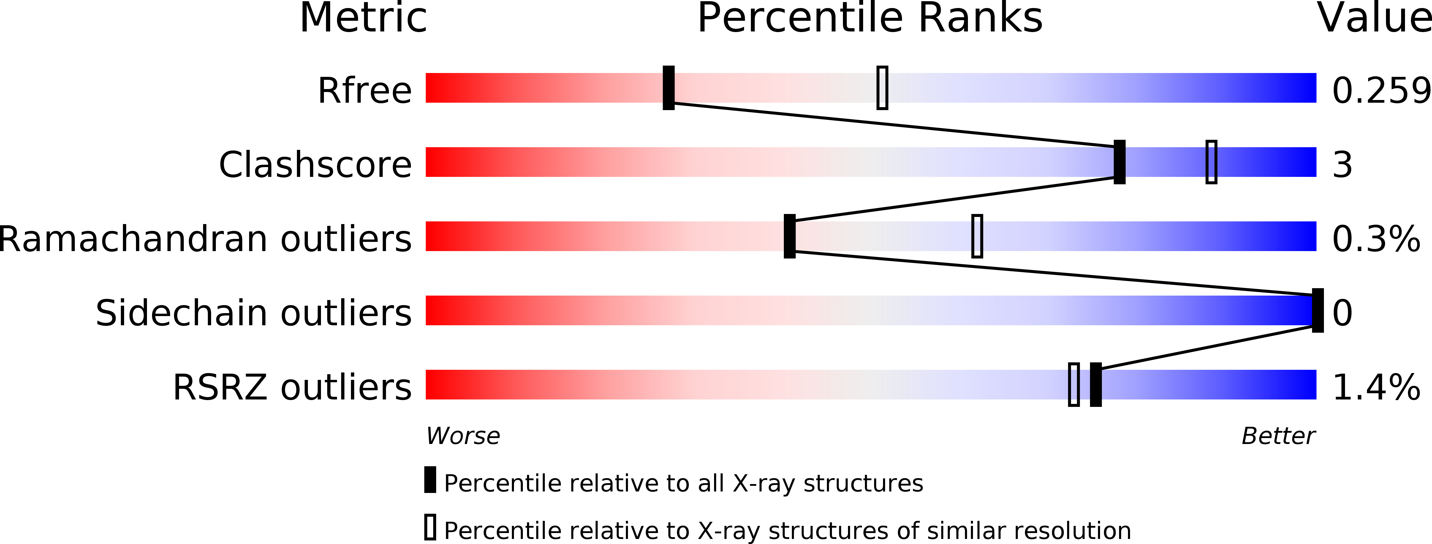
Resolution:
2.58 Å
R-Value Free:
0.25
R-Value Work:
0.19
R-Value Observed:
0.19
Space Group:
C 1 2 1
Macromolecular Entities
Structures with similar UniProt ID
Protein Blast
Polymer Type:polypeptide(L)
Molecule:Dual specificity tyrosine-phosphorylation-regulated kinase 2
Gene (Uniprot):DYRK2
Chain IDs:A, B
Chain Length:408
Number of Molecules:2
Biological Source:Homo sapiens
Modified Residue
Compound ID Chain ID Parent Comp ID Details 2D Image
PTR A TYR modified residue
Primary Citation
An Unusual Binding Model of the Methyl 9-Anilinothiazolo[5,4-f] quinazoline-2-carbimidates (EHT 1610 and EHT 5372) Confers High Selectivity for Dual-Specificity Tyrosine Phosphorylation-Regulated Kinases.
J. Med. Chem. 59 10315 10321 (2016)
PMID: 27766861 DOI: 10.1021/acs.jmedchem.6b01083

Abstact

Methyl 9-anilinothiazolo[5,4-f]quinazoline-2-carbimidates 1 (EHT 5372) and 2 (EHT 1610) are strong inhibitors of DYRK's family kinases. The crystal structures of the complex revealed a noncanonical binding mode of compounds 1 and 2 in DYRK2, explaining the remarkable selectivity and potency of these inhibitors. The structural data and comparison presented here provide therefore a template for further improvement of this inhibitor class and for the development of novel inhibitors selectively targeting DYRK kinases.

Legend

Protein

Chemical

Disease

Primary Citation of related structures
Feedback Form
Name
Email
Institute
Feedback