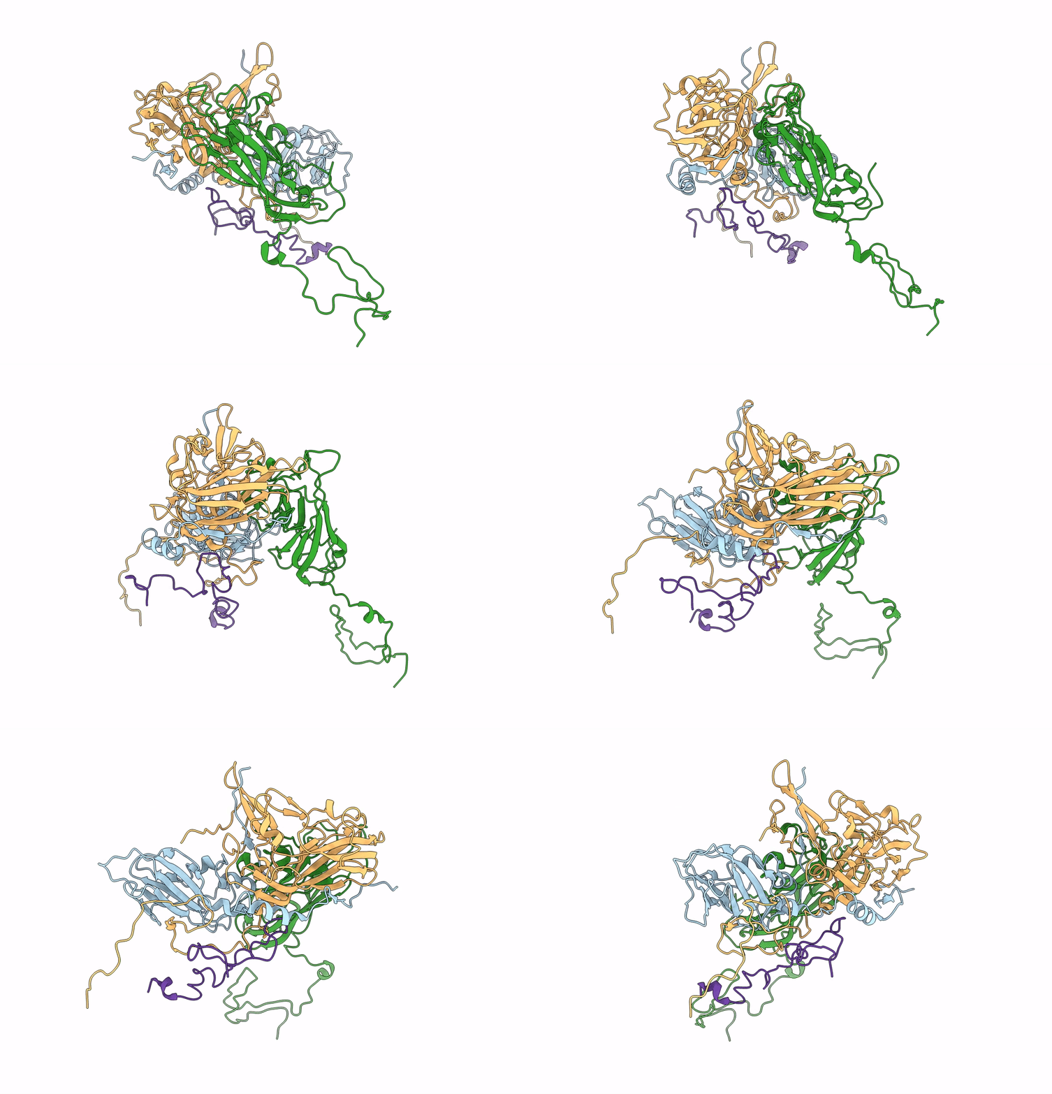
5LWG image
Deposition Date 2016-09-16
Release Date 2016-11-30
Last Version Date 2024-05-15
Entry Detail
PDB ID:
5LWG
Keywords:
Title:
Israeli acute paralysis virus heated to 63 degree - full particle
Biological Source:
Source Organism:
Method Details:
Experimental Method:
Resolution:
3.20 Å
Aggregation State:
PARTICLE
Reconstruction Method:
SINGLE PARTICLE
Macromolecular Entities
Polymer Type:polypeptide(L)
Molecule:VP1
Chain IDs:A
Chain Length:208
Number of Molecules:1
Biological Source:Israeli acute paralysis virus
Polymer Type:polypeptide(L)
Molecule:VP2
Chain IDs:C (auth: B)
Chain Length:247
Number of Molecules:1
Biological Source:Israeli acute paralysis virus
Polymer Type:polypeptide(L)
Molecule:VP3
Chain IDs:B (auth: C)
Chain Length:300
Number of Molecules:1
Biological Source:Israeli acute paralysis virus
Polymer Type:polypeptide(L)
Molecule:VP4
Chain IDs:D
Chain Length:57
Number of Molecules:1
Biological Source:Israeli acute paralysis virus
Ligand Molecules
Primary Citation
Cryo-electron Microscopy Study of the Genome Release of the Dicistrovirus Israeli Acute Bee Paralysis Virus.
J. Virol. 91 ? ? (2017)
PMID: 27928006 DOI: 10.1128/JVI.02060-16

Abstact

UNLABELLED Viruses of the family Dicistroviridae can cause substantial economic damage by infecting agriculturally important insects. Israeli acute bee paralysis virus (IAPV) causes honeybee colony collapse disorder in the United States. High-resolution molecular details of the genome delivery mechanism of dicistroviruses are unknown. Here we present a cryo-electron microscopy analysis of IAPV virions induced to release their genomes in vitro We determined structures of full IAPV virions primed to release their genomes to a resolution of 3.3 Å and of empty capsids to a resolution of 3.9 Å. We show that IAPV does not form expanded A particles before genome release as in the case of related enteroviruses of the family Picornaviridae The structural changes observed in the empty IAPV particles include detachment of the VP4 minor capsid proteins from the inner face of the capsid and partial loss of the structure of the N-terminal arms of the VP2 capsid proteins. Unlike the case for many picornaviruses, the empty particles of IAPV are not expanded relative to the native virions and do not contain pores in their capsids that might serve as channels for genome release. Therefore, rearrangement of a unique region of the capsid is probably required for IAPV genome release. IMPORTANCE Honeybee populations in Europe and North America are declining due to pressure from pathogens, including viruses. Israeli acute bee paralysis virus (IAPV), a member of the family Dicistroviridae, causes honeybee colony collapse disorder in the United States. The delivery of virus genomes into host cells is necessary for the initiation of infection. Here we present a structural cryo-electron microscopy analysis of IAPV particles induced to release their genomes. We show that genome release is not preceded by an expansion of IAPV virions as in the case of related picornaviruses that infect vertebrates. Furthermore, minor capsid proteins detach from the capsid upon genome release. The genome leaves behind empty particles that have compact protein shells.

Legend

Protein

Chemical

Disease

Primary Citation of related structures