Deposition Date
2016-09-12
Release Date
2017-02-22
Last Version Date
2024-01-17
Entry Detail
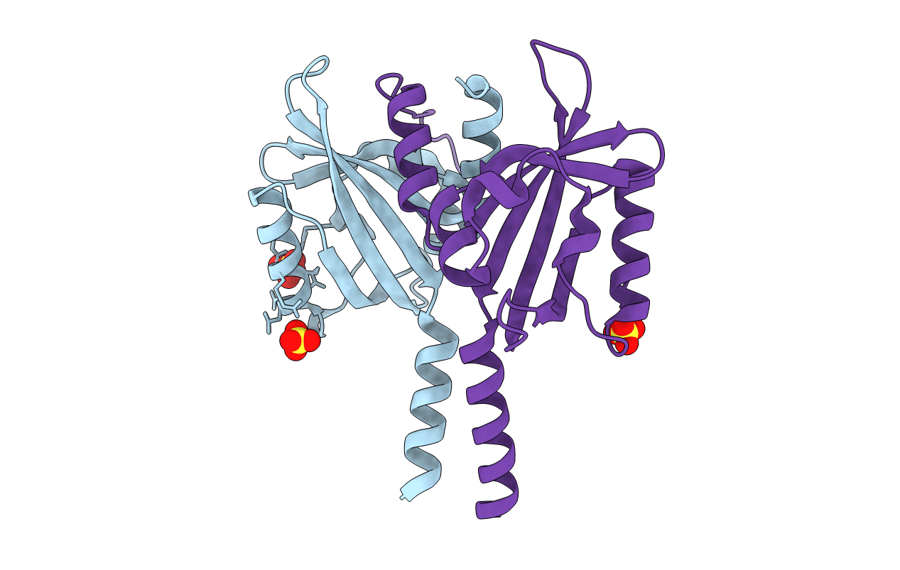
Biological Source:
Source Organism(s):
Pseudomonas putida (Taxon ID: 390235)
Expression System(s):
Method Details:
Experimental Method:
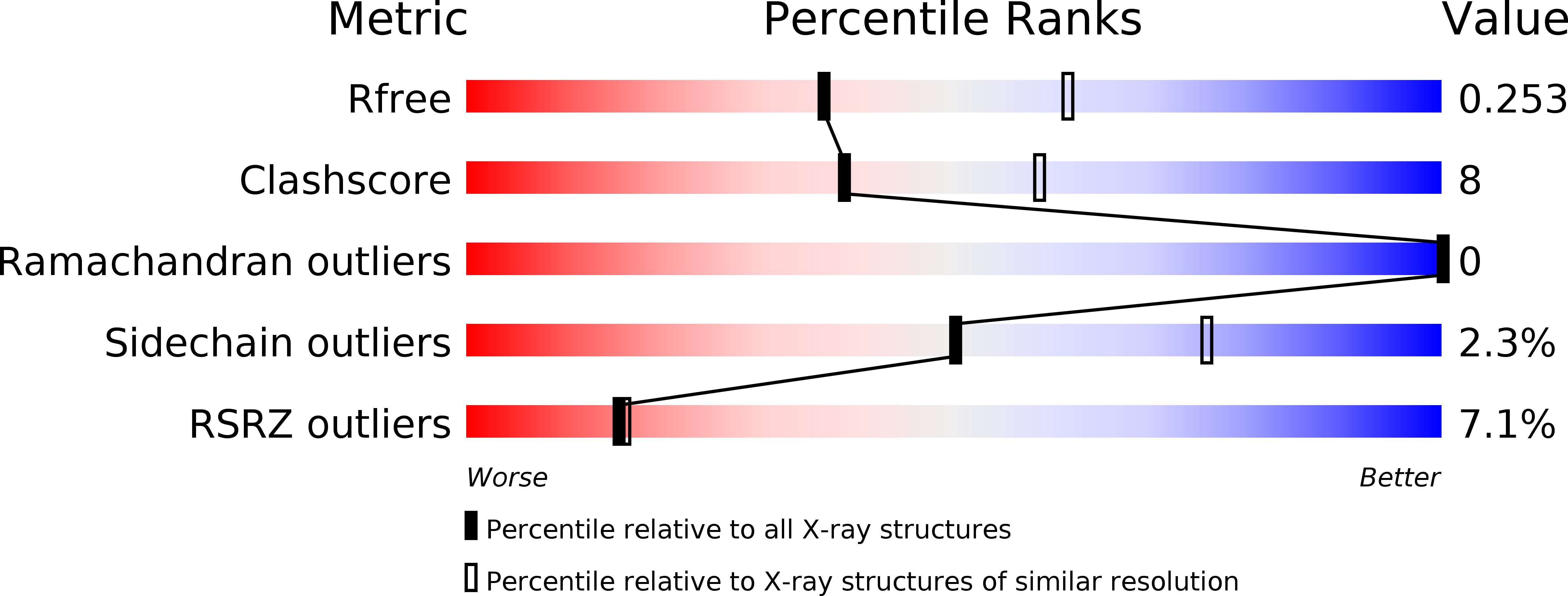
Resolution:
2.50 Å
R-Value Free:
0.25
R-Value Work:
0.21
R-Value Observed:
0.21
Space Group:
I 41